4KA9

Crystal structure analysis of single amino acid deletion mutations in EGFP


Experimental Data Snapshot

  • Method: X-RAY DIFFRACTION
  • Resolution: 1.58 Å
  • R-Value Free: 0.210 
  • R-Value Work: 0.173 
  • R-Value Observed: 0.175 

wwPDB Validation   3D Report Full Report


This is version 1.1 of the entry. See complete history


Literature

Random single amino acid deletion sampling unveils structural tolerance and the benefits of helical registry shift on GFP folding and structure.

Arpino, J.A.Reddington, S.C.Halliwell, L.M.Rizkallah, P.J.Jones, D.D.

(2014) Structure 22: 889-898

  • DOI: https://doi.org/10.1016/j.str.2014.03.014
  • Primary Citation of Related Structures:  
    4KA9

  • PubMed Abstract: 

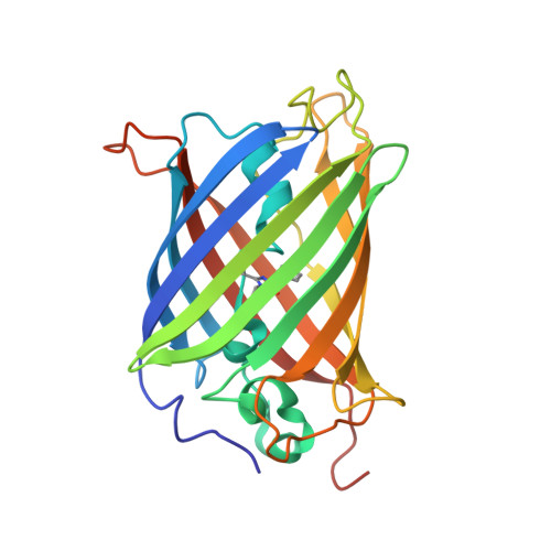
    Altering a protein's backbone through amino acid deletion is a common evolutionary mutational mechanism, but is generally ignored during protein engineering primarily because its effect on the folding-structure-function relationship is difficult to predict. Using directed evolution, enhanced green fluorescent protein (EGFP) was observed to tolerate residue deletion across the breadth of the protein, particularly within short and long loops, helical elements, and at the termini of strands. A variant with G4 removed from a helix (EGFP(G4Δ)) conferred significantly higher cellular fluorescence. Folding analysis revealed that EGFP(G4Δ) retained more structure upon unfolding and refolded with almost 100% efficiency but at the expense of thermodynamic stability. The EGFP(G4Δ) structure revealed that G4 deletion caused a beneficial helical registry shift resulting in a new polar interaction network, which potentially stabilizes a cis proline peptide bond and links secondary structure elements. Thus, deletion mutations and registry shifts can enhance proteins through structural rearrangements not possible by substitution mutations alone.


  • Organizational Affiliation

    School of Biosciences, Main Building, Park Place, Cardiff University, Cardiff CF10 3AT, UK.


Macromolecules
Find similar proteins by:  (by identity cutoff)  |  3D Structure
Entity ID: 1
MoleculeChains Sequence LengthOrganismDetailsImage
Green fluorescent protein236Aequorea victoriaMutation(s): 2 
Gene Names: GFP
UniProt
Find proteins for P42212 (Aequorea victoria)
Explore P42212 
Go to UniProtKB:  P42212
Entity Groups  
Sequence Clusters30% Identity50% Identity70% Identity90% Identity95% Identity100% Identity
UniProt GroupP42212
Sequence Annotations
Expand
  • Reference Sequence
Small Molecules
Ligands 4 Unique
IDChains Name / Formula / InChI Key2D Diagram3D Interactions
EPE
Query on EPE

Download Ideal Coordinates CCD File 
K [auth A]4-(2-HYDROXYETHYL)-1-PIPERAZINE ETHANESULFONIC ACID
C8 H18 N2 O4 S
JKMHFZQWWAIEOD-UHFFFAOYSA-N
ZN
Query on ZN

Download Ideal Coordinates CCD File 
M [auth A],
N [auth A]
ZINC ION
Zn
PTFCDOFLOPIGGS-UHFFFAOYSA-N
EDO
Query on EDO

Download Ideal Coordinates CCD File 
B [auth A]
C [auth A]
D [auth A]
E [auth A]
F [auth A]
B [auth A],
C [auth A],
D [auth A],
E [auth A],
F [auth A],
G [auth A],
H [auth A],
I [auth A],
J [auth A]
1,2-ETHANEDIOL
C2 H6 O2
LYCAIKOWRPUZTN-UHFFFAOYSA-N
NA
Query on NA

Download Ideal Coordinates CCD File 
L [auth A],
O [auth A]
SODIUM ION
Na
FKNQFGJONOIPTF-UHFFFAOYSA-N
Modified Residues  1 Unique
IDChains TypeFormula2D DiagramParent
CRO
Query on CRO
A
L-PEPTIDE LINKINGC15 H17 N3 O5THR, TYR, GLY
Experimental Data & Validation

Experimental Data

  • Method: X-RAY DIFFRACTION
  • Resolution: 1.58 Å
  • R-Value Free: 0.210 
  • R-Value Work: 0.173 
  • R-Value Observed: 0.175 
  • Space Group: C 1 2 1
Unit Cell:
Length ( Å )Angle ( ˚ )
a = 91.87α = 90
b = 66.66β = 108.76
c = 45.29γ = 90
Software Package:
Software NamePurpose
SCALAdata scaling
PHASERphasing
REFMACrefinement
PDB_EXTRACTdata extraction

Structure Validation

View Full Validation Report


Entry History 

Deposition Data

Revision History  (Full details and data files)

  • Version 1.0: 2014-05-07
    Type: Initial release
  • Version 1.1: 2015-11-25
    Changes: Database references, Structure summary