4KI8

Crystal structure of a GroEL-ADP complex in the relaxed allosteric state


Experimental Data Snapshot

  • Method: X-RAY DIFFRACTION
  • Resolution: 2.72 Å
  • R-Value Free: 0.203 
  • R-Value Work: 0.166 
  • R-Value Observed: 0.166 

wwPDB Validation   3D Report Full Report


Ligand Structure Quality Assessment 


This is version 1.5 of the entry. See complete history


Literature

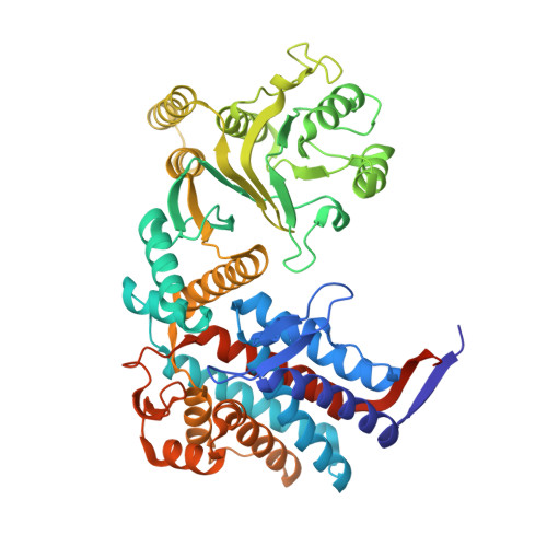
Crystal structure of a GroEL-ADP complex in the relaxed allosteric state at 2.7 A resolution.

Fei, X.Yang, D.Laronde-Leblanc, N.Lorimer, G.H.

(2013) Proc Natl Acad Sci U S A 110: E2958-E2966

  • DOI: https://doi.org/10.1073/pnas.1311996110
  • Primary Citation of Related Structures:  
    4KI8

  • PubMed Abstract: 

    The chaperonin proteins GroEL and GroES are cellular nanomachines driven by the hydrolysis of ATP that facilitate the folding of structurally diverse substrate proteins. In response to ligand binding, the subunits of a ring cycle in a concerted manner through a series of allosteric states (T, R, and R″), enabling work to be performed on the substrate protein. Removing two salt bridges that ordinarily break during the allosteric transitions of the WT permitted the structure of GroEL-ADP in the R state to be solved to 2.7 Å resolution. Whereas the equatorial domain displays almost perfect sevenfold symmetry, the apical domains, to which substrate proteins bind, and to a lesser extent, the intermediate domains display a remarkable asymmetry. Freed of intersubunit contacts, the apical domain of each subunit adopts a different conformation, suggesting a flexibility that permits interaction with diverse substrate proteins. This result contrasts with a previous cryo-EM study of a related allosteric ATP-bound state at lower resolution. After artificially imposing sevenfold symmetry it was concluded that a GroEL ring in the R-ATP state existed in six homogeneous but slightly different states. By imposing sevenfold symmetry on each of the subunits of the crystal structure of GroEL-ADP, we showed that the synthetic rings of (X-ray) GroEL-ADP and (cryo-EM) GroEL-ATP are structurally closely related. A deterministic model, the click stop mechanism, that implied temporal transitions between these states was proposed. Here, however, these conformational states are shown to exist as a structurally heterogeneous ensemble within a single ring.


  • Organizational Affiliation

    Center for Biological Structure and Organization, University of Maryland, College Park, MD 20742, USA.


Macromolecules
Find similar proteins by:  (by identity cutoff)  |  3D Structure
Entity ID: 1
MoleculeChains Sequence LengthOrganismDetailsImage
GroEL protein
A, B, C, D, E
A, B, C, D, E, F, G
548Escherichia coliMutation(s): 2 
Gene Names: BN17_41231ECs5124groELgroLLF82_0923mopA
UniProt
Find proteins for Q548M1 (Escherichia coli)
Explore Q548M1 
Go to UniProtKB:  Q548M1
Entity Groups  
Sequence Clusters30% Identity50% Identity70% Identity90% Identity95% Identity100% Identity
UniProt GroupQ548M1
Sequence Annotations
Expand
  • Reference Sequence
Small Molecules
Ligands 5 Unique
IDChains Name / Formula / InChI Key2D Diagram3D Interactions
ADP
Query on ADP

Download Ideal Coordinates CCD File 
BA [auth C]
FA [auth D]
H [auth A]
MA [auth E]
S [auth B]
BA [auth C],
FA [auth D],
H [auth A],
MA [auth E],
S [auth B],
TA [auth F],
ZA [auth G]
ADENOSINE-5'-DIPHOSPHATE
C10 H15 N5 O10 P2
XTWYTFMLZFPYCI-KQYNXXCUSA-N
MPD
Query on MPD

Download Ideal Coordinates CCD File 
CB [auth G]
DB [auth G]
EA [auth C]
IA [auth D]
JA [auth D]
CB [auth G],
DB [auth G],
EA [auth C],
IA [auth D],
JA [auth D],
K [auth A],
KA [auth D],
L [auth A],
M [auth A],
N [auth A],
O [auth A],
P [auth A],
PA [auth E],
V [auth B],
W [auth B],
WA [auth F],
X [auth B],
Y [auth B],
Z [auth B]
(4S)-2-METHYL-2,4-PENTANEDIOL
C6 H14 O2
SVTBMSDMJJWYQN-YFKPBYRVSA-N
CA
Query on CA

Download Ideal Coordinates CCD File 
AA [auth B]
EB [auth G]
FB [auth G]
HB [auth G]
LA [auth D]
AA [auth B],
EB [auth G],
FB [auth G],
HB [auth G],
LA [auth D],
Q [auth A],
QA [auth E],
R [auth A],
RA [auth E],
SA [auth E],
YA [auth F]
CALCIUM ION
Ca
BHPQYMZQTOCNFJ-UHFFFAOYSA-N
K
Query on K

Download Ideal Coordinates CCD File 
BB [auth G]
DA [auth C]
GB [auth G]
HA [auth D]
J [auth A]
BB [auth G],
DA [auth C],
GB [auth G],
HA [auth D],
J [auth A],
OA [auth E],
U [auth B],
VA [auth F],
XA [auth F]
POTASSIUM ION
K
NPYPAHLBTDXSSS-UHFFFAOYSA-N
MG
Query on MG

Download Ideal Coordinates CCD File 
AB [auth G]
CA [auth C]
GA [auth D]
I [auth A]
NA [auth E]
AB [auth G],
CA [auth C],
GA [auth D],
I [auth A],
NA [auth E],
T [auth B],
UA [auth F]
MAGNESIUM ION
Mg
JLVVSXFLKOJNIY-UHFFFAOYSA-N
Experimental Data & Validation

Experimental Data

  • Method: X-RAY DIFFRACTION
  • Resolution: 2.72 Å
  • R-Value Free: 0.203 
  • R-Value Work: 0.166 
  • R-Value Observed: 0.166 
  • Space Group: C 1 2 1
Unit Cell:
Length ( Å )Angle ( ˚ )
a = 235.222α = 90
b = 141.655β = 113.84
c = 156.693γ = 90
Software Package:
Software NamePurpose
SERGUIdata collection
PHASESphasing
PHENIXrefinement
HKL-2000data reduction
HKL-2000data scaling

Structure Validation

View Full Validation Report


Ligand Structure Quality Assessment 


Entry History 

Deposition Data

Revision History  (Full details and data files)

  • Version 1.0: 2013-07-17
    Type: Initial release
  • Version 1.1: 2013-08-07
    Changes: Database references
  • Version 1.2: 2013-08-14
    Changes: Other
  • Version 1.3: 2013-08-28
    Changes: Database references
  • Version 1.4: 2013-11-13
    Changes: Other
  • Version 1.5: 2023-09-20
    Changes: Data collection, Database references, Derived calculations, Refinement description