4KU1

Role of the hinge and C-gamma-2/C-gamma-3 interface in immunoglobin G1 Fc domain motions: implications for Fc engineering


Experimental Data Snapshot

  • Method: X-RAY DIFFRACTION
  • Resolution: 1.90 Å
  • R-Value Free: 0.258 
  • R-Value Work: 0.212 
  • R-Value Observed: 0.215 

wwPDB Validation   3D Report Full Report


This is version 2.0 of the entry. See complete history


Literature

Immunoglobulin g1 fc domain motions: implications for fc engineering.

Frank, M.Walker, R.C.Lanzilotta, W.N.Prestegard, J.H.Barb, A.W.

(2014) J Mol Biol 426: 1799-1811

  • DOI: https://doi.org/10.1016/j.jmb.2014.01.011
  • Primary Citation of Related Structures:  
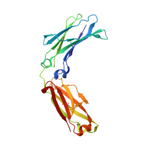
    4KU1

  • PubMed Abstract: 

    The fragment crystallizable (Fc) region links the key pathogen identification and destruction properties of immunoglobulin G (IgG). Pathogen opsonization positions Fcs to activate pro-inflammatory Fcγ receptors (FcγRs) on immune cells. The cellular response and committal to a damaging, though protective, immune response are tightly controlled at multiple levels. Control mechanisms are diverse and in many cases unclear, but one frequently suggested contribution originates in FcγR affinity being modulated through shifts in Fc conformational sampling. Here, we report a previously unseen IgG1 Fc conformation. This observation motivated an extensive molecular dynamics investigation of polypeptide and glycan motions that revealed greater amplitude of motion for the N-terminal Cγ2 domains and N-glycan than previously observed. Residues in the Cγ2/Cγ3 interface and disulfide-bonded hinge were identified as influencing the Cγ2 motion. Our results are consistent with a model of Fc that is structurally dynamic. Conformational states that are competent to bind immune-stimulating FcγRs interconverted with Fc conformations distinct from those observed in FcγR complexes, which may represent a transient, nonbinding population.


  • Organizational Affiliation

    Biognos AB, Generatorsgatan 1, 41705 Gothenburg, Sweden.


Macromolecules
Find similar proteins by:  (by identity cutoff)  |  3D Structure
Entity ID: 1
MoleculeChains Sequence LengthOrganismDetailsImage
Ig gamma-1 chain C region
A, B
208Homo sapiensMutation(s): 0 
UniProt & NIH Common Fund Data Resources
Find proteins for P01857 (Homo sapiens)
Explore P01857 
Go to UniProtKB:  P01857
PHAROS:  P01857
GTEx:  ENSG00000211896 
Entity Groups  
Sequence Clusters30% Identity50% Identity70% Identity90% Identity95% Identity100% Identity
UniProt GroupP01857
Sequence Annotations
Expand
  • Reference Sequence
Oligosaccharides

Help

Entity ID: 2
MoleculeChains Length2D Diagram Glycosylation3D Interactions
beta-D-galactopyranose-(1-4)-2-acetamido-2-deoxy-beta-D-glucopyranose-(1-2)-alpha-D-mannopyranose-(1-3)-[beta-D-galactopyranose-(1-4)-2-acetamido-2-deoxy-beta-D-glucopyranose-(1-2)-alpha-D-mannopyranose-(1-6)]beta-D-mannopyranose-(1-4)-2-acetamido-2-deoxy-beta-D-glucopyranose-(1-4)-[beta-L-fucopyranose-(1-6)]2-acetamido-2-deoxy-beta-D-glucopyranose
C
10N-Glycosylation
Glycosylation Resources
GlyTouCan:  G23770JR
GlyCosmos:  G23770JR
GlyGen:  G23770JR
Entity ID: 3
MoleculeChains Length2D Diagram Glycosylation3D Interactions
beta-D-galactopyranose-(1-4)-2-acetamido-2-deoxy-beta-D-glucopyranose-(1-2)-alpha-D-mannopyranose-(1-6)-[2-acetamido-2-deoxy-beta-D-glucopyranose-(1-2)-alpha-D-mannopyranose-(1-3)]beta-D-mannopyranose-(1-4)-2-acetamido-2-deoxy-beta-D-glucopyranose-(1-4)-[beta-L-fucopyranose-(1-6)]2-acetamido-2-deoxy-beta-D-glucopyranose
D
9N-Glycosylation
Glycosylation Resources
GlyTouCan:  G33244HE
GlyCosmos:  G33244HE
GlyGen:  G33244HE
Small Molecules
Ligands 1 Unique
IDChains Name / Formula / InChI Key2D Diagram3D Interactions
PG4
Query on PG4

Download Ideal Coordinates CCD File 
E [auth A]TETRAETHYLENE GLYCOL
C8 H18 O5
UWHCKJMYHZGTIT-UHFFFAOYSA-N
Experimental Data & Validation

Experimental Data

  • Method: X-RAY DIFFRACTION
  • Resolution: 1.90 Å
  • R-Value Free: 0.258 
  • R-Value Work: 0.212 
  • R-Value Observed: 0.215 
  • Space Group: P 21 21 21
Unit Cell:
Length ( Å )Angle ( ˚ )
a = 49.145α = 90
b = 79.865β = 90
c = 128.402γ = 90
Software Package:
Software NamePurpose
REFMACrefinement

Structure Validation

View Full Validation Report


Entry History 

Deposition Data

Revision History  (Full details and data files)

  • Version 1.0: 2014-02-19
    Type: Initial release
  • Version 1.1: 2014-03-19
    Changes: Database references
  • Version 1.2: 2014-04-09
    Changes: Database references
  • Version 2.0: 2020-07-29
    Type: Remediation
    Reason: Carbohydrate remediation
    Changes: Advisory, Atomic model, Data collection, Derived calculations, Structure summary