4M7J

Crystal structure of S25-26 in complex with Kdo(2.8)Kdo(2.4)Kdo trisaccharide


Experimental Data Snapshot

  • Method: X-RAY DIFFRACTION
  • Resolution: 1.95 Å
  • R-Value Free: 0.247 
  • R-Value Work: 0.212 
  • R-Value Observed: 0.213 

wwPDB Validation   3D Report Full Report


Ligand Structure Quality Assessment 


This is version 2.1 of the entry. See complete history


Literature

Groove-type Recognition of Chlamydiaceae-specific Lipopolysaccharide Antigen by a Family of Antibodies Possessing an Unusual Variable Heavy Chain N-Linked Glycan.

Haji-Ghassemi, O.Muller-Loennies, S.Saldova, R.Muniyappa, M.Brade, L.Rudd, P.M.Harvey, D.J.Kosma, P.Brade, H.Evans, S.V.

(2014) J Biol Chem 289: 16644-16661

  • DOI: https://doi.org/10.1074/jbc.M113.528224
  • Primary Citation of Related Structures:  
    4M7J, 4M7Z, 4M93, 4MA1

  • PubMed Abstract: 

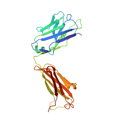
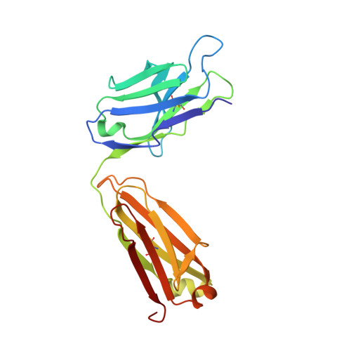
    The structure of the antigen binding fragment of mAb S25-26, determined to 1.95 Å resolution in complex with the Chlamydiaceae family-specific trisaccharide antigen Kdo(2→8)Kdo(2→4)Kdo (Kdo = 3-deoxy-α-d-manno-oct-2-ulopyranosonic acid), displays a germ-line-coded paratope that differs significantly from previously characterized Chlamydiaceae-specific mAbs despite being raised against the identical immunogen. Unlike the terminal Kdo recognition pocket that promotes cross-reactivity in S25-2-type antibodies, S25-26 and the closely related S25-23 utilize a groove composed of germ-line residues to recognize the entire trisaccharide antigen and so confer strict specificity. Interest in S25-23 was sparked by its rare high μm affinity and strict specificity for the family-specific trisaccharide antigen; however, only the related antibody S25-26 proved amenable to crystallization. The structures of three unliganded forms of S25-26 have a labile complementary-determining region H3 adjacent to significant glycosylation of the variable heavy chain on asparagine 85 in Framework Region 3. Analysis of the glycan reveals a heterogeneous mixture with a common root structure that contains an unusually high number of terminal αGal-Gal moieties. One of the few reported structures of glycosylated mAbs containing these epitopes is the therapeutic antibody Cetuximab; however, unlike Cetuximab, one of the unliganded structures in S25-26 shows significant order in the glycan with appropriate electron density for nine residues. The elucidation of the three-dimensional structure of an αGal-containing N-linked glycan on a mAb variable heavy chain has potential clinical interest, as it has been implicated in allergic response in patients receiving therapeutic antibodies.


  • Organizational Affiliation

    From the Department of Biochemistry and Microbiology, University of Victoria, Victoria, British Columbia V8P 3P6, Canada.


Macromolecules
Find similar proteins by:  (by identity cutoff)  |  3D Structure
Entity ID: 1
MoleculeChains Sequence LengthOrganismDetailsImage
S25-26 Fab (IgG1k) heavy chainA [auth H]219Mus musculusMutation(s): 0 
Entity Groups  
Sequence Clusters30% Identity50% Identity70% Identity90% Identity95% Identity100% Identity
Sequence Annotations
Expand
  • Reference Sequence
Find similar proteins by:  (by identity cutoff)  |  3D Structure
Entity ID: 2
MoleculeChains Sequence LengthOrganismDetailsImage
S25-26 Fab (Igg1k) light chainB [auth L]219Mus musculusMutation(s): 0 
Entity Groups  
Sequence Clusters30% Identity50% Identity70% Identity90% Identity95% Identity100% Identity
Sequence Annotations
Expand
  • Reference Sequence
Oligosaccharides

Help

Entity ID: 3
MoleculeChains Length2D Diagram Glycosylation3D Interactions
3-deoxy-alpha-D-manno-oct-2-ulopyranosonic acid-(2-8)-3-deoxy-alpha-D-manno-oct-2-ulopyranosonic acid-(2-4)-prop-2-en-1-yl 3-deoxy-alpha-D-manno-oct-2-ulopyranosidonic acidC [auth A]3N/AN/A
Glycosylation Resources
GlyTouCan:  G25386BJ
GlyCosmos:  G25386BJ
Small Molecules
Experimental Data & Validation

Experimental Data

  • Method: X-RAY DIFFRACTION
  • Resolution: 1.95 Å
  • R-Value Free: 0.247 
  • R-Value Work: 0.212 
  • R-Value Observed: 0.213 
  • Space Group: P 32 2 1
Unit Cell:
Length ( Å )Angle ( ˚ )
a = 74.464α = 90
b = 74.464β = 90
c = 149.893γ = 120
Software Package:
Software NamePurpose
DENZOdata reduction
SCALEPACKdata scaling
PHASERphasing
REFMACrefinement
PDB_EXTRACTdata extraction
Macromoleculardata collection

Structure Validation

View Full Validation Report


Ligand Structure Quality Assessment 


Entry History 

Deposition Data

Revision History  (Full details and data files)

  • Version 1.0: 2014-04-09
    Type: Initial release
  • Version 1.1: 2014-06-11
    Changes: Database references
  • Version 1.2: 2014-07-09
    Changes: Database references
  • Version 1.3: 2017-11-15
    Changes: Refinement description
  • Version 2.0: 2020-07-29
    Type: Remediation
    Reason: Carbohydrate remediation
    Changes: Atomic model, Data collection, Derived calculations, Structure summary
  • Version 2.1: 2023-09-20
    Changes: Data collection, Database references, Refinement description, Structure summary