4PL8

Structure of rabbit skeletal muscle actin in complex with a hybrid peptide comprising thymosin beta4 and the lysine-rich region of Cordon-Bleu


Experimental Data Snapshot

  • Method: X-RAY DIFFRACTION
  • Resolution: 2.00 Å
  • R-Value Free: 0.207 
  • R-Value Work: 0.171 
  • R-Value Observed: 0.173 

wwPDB Validation   3D Report Full Report


Ligand Structure Quality Assessment 


This is version 1.2 of the entry. See complete history


Literature

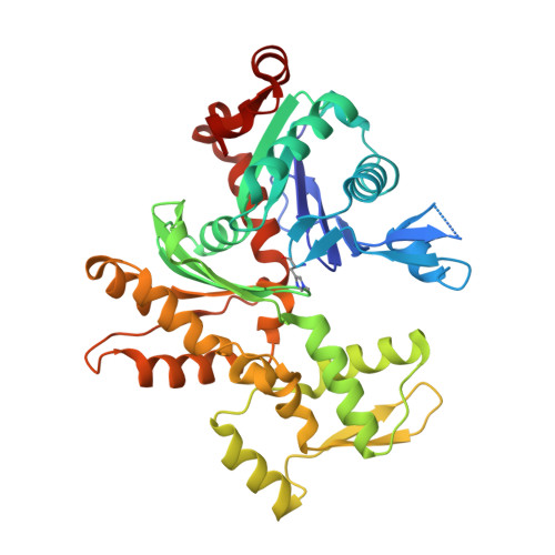
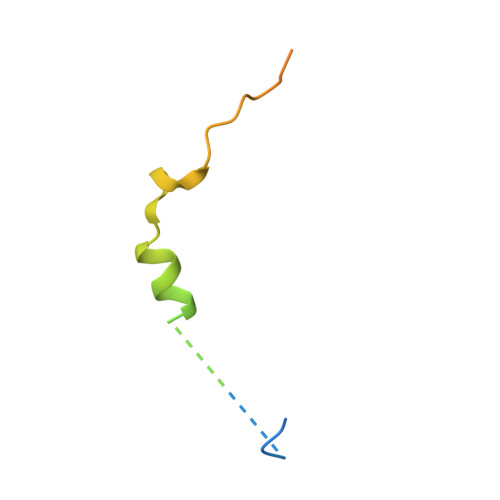
Structural basis of thymosin-beta 4/profilin exchange leading to actin filament polymerization.

Xue, B.Leyrat, C.Grimes, J.M.Robinson, R.C.

(2014) Proc Natl Acad Sci U S A 111: E4596-E4605

  • DOI: https://doi.org/10.1073/pnas.1412271111
  • Primary Citation of Related Structures:  
    4PL7, 4PL8

  • PubMed Abstract: 

    Thymosin-β4 (Tβ4) and profilin are the two major sequestering proteins that maintain the pool of monomeric actin (G-actin) within cells of higher eukaryotes. Tβ4 prevents G-actin from joining a filament, whereas profilin:actin only supports barbed-end elongation. Here, we report two Tβ4:actin structures. The first structure shows that Tβ4 has two helices that bind at the barbed and pointed faces of G-actin, preventing the incorporation of the bound G-actin into a filament. The second structure displays a more open nucleotide binding cleft on G-actin, which is typical of profilin:actin structures, with a concomitant disruption of the Tβ4 C-terminal helix interaction. These structures, combined with biochemical assays and molecular dynamics simulations, show that the exchange of bound actin between Tβ4 and profilin involves both steric and allosteric components. The sensitivity of profilin to the conformational state of actin indicates a similar allosteric mechanism for the dissociation of profilin during filament elongation.


  • Organizational Affiliation

    Institute of Molecular and Cell Biology, A*STAR (Agency for Science, Technology and Research), Biopolis, Singapore 138673; bxue@imcb.a-star.edu.sg.


Macromolecules
Find similar proteins by:  (by identity cutoff)  |  3D Structure
Entity ID: 1
MoleculeChains Sequence LengthOrganismDetailsImage
Actin, alpha skeletal muscleA,
C [auth B]
375Oryctolagus cuniculusMutation(s): 0 
UniProt
Find proteins for P68135 (Oryctolagus cuniculus)
Explore P68135 
Go to UniProtKB:  P68135
Entity Groups  
Sequence Clusters30% Identity50% Identity70% Identity90% Identity95% Identity100% Identity
UniProt GroupP68135
Sequence Annotations
Expand
  • Reference Sequence
Find similar proteins by:  (by identity cutoff)  |  3D Structure
Entity ID: 2
MoleculeChains Sequence LengthOrganismDetailsImage
Thymosin beta-4,Protein cordon-bleu,Thymosin beta-4B [auth H]73Homo sapiensMutation(s): 0 
UniProt & NIH Common Fund Data Resources
Find proteins for O75128 (Homo sapiens)
Explore O75128 
Go to UniProtKB:  O75128
PHAROS:  O75128
GTEx:  ENSG00000106078 
Find proteins for P62328 (Homo sapiens)
Explore P62328 
Go to UniProtKB:  P62328
PHAROS:  P62328
GTEx:  ENSG00000205542 
Entity Groups  
Sequence Clusters30% Identity50% Identity70% Identity90% Identity95% Identity100% Identity
UniProt GroupsP62328O75128
Sequence Annotations
Expand
  • Reference Sequence
Small Molecules
Experimental Data & Validation

Experimental Data

  • Method: X-RAY DIFFRACTION
  • Resolution: 2.00 Å
  • R-Value Free: 0.207 
  • R-Value Work: 0.171 
  • R-Value Observed: 0.173 
  • Space Group: P 43 21 2
Unit Cell:
Length ( Å )Angle ( ˚ )
a = 93.64α = 90
b = 93.64β = 90
c = 206.274γ = 90
Software Package:
Software NamePurpose
DENZOdata collection
SCALEPACKdata reduction
PHASERphasing
PDB_EXTRACTdata extraction
Cootmodel building
PHENIXrefinement
PHASERmodel building

Structure Validation

View Full Validation Report


Ligand Structure Quality Assessment 


Entry History 

Deposition Data

Revision History  (Full details and data files)

  • Version 1.0: 2014-10-22
    Type: Initial release
  • Version 1.1: 2014-11-12
    Changes: Database references
  • Version 1.2: 2023-11-08
    Changes: Data collection, Database references, Derived calculations, Other, Refinement description, Source and taxonomy