4QXG

Antigen binding fragment of an anti IFNAR1 antibody


Experimental Data Snapshot

  • Method: X-RAY DIFFRACTION
  • Resolution: 2.30 Å
  • R-Value Free: 0.249 
  • R-Value Work: 0.190 
  • R-Value Observed: 0.193 

wwPDB Validation   3D Report Full Report


This is version 1.1 of the entry. See complete history


Literature

Molecular basis for antagonistic activity of anifrolumab, an anti-interferon-alpha receptor 1 antibody.

Peng, L.Oganesyan, V.Wu, H.Dall'Acqua, W.F.Damschroder, M.M.

(2015) MAbs 7: 428-439

  • DOI: https://doi.org/10.1080/19420862.2015.1007810
  • Primary Citation of Related Structures:  
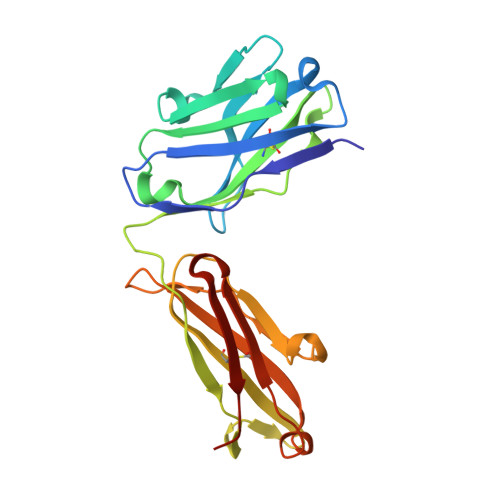
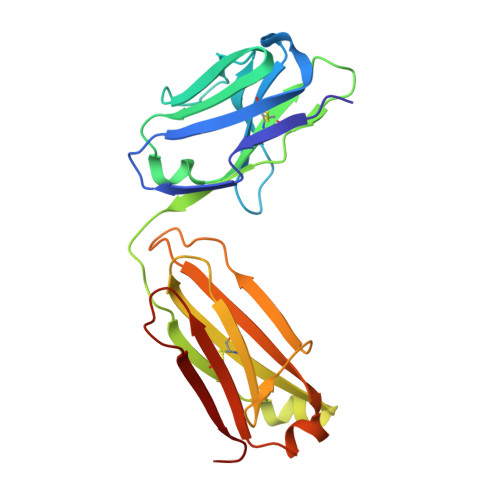
    4QXG

  • PubMed Abstract: 

    Anifrolumab (anifrolumab) is an antagonist human monoclonal antibody that targets interferon α receptor 1 (IFNAR1). Anifrolumab has been developed to treat autoimmune diseases and is currently in clinical trials. To decipher the molecular basis of its mechanism of action, we engaged in multiple epitope mapping approaches to determine how it interacts with IFNAR1 and antagonizes the receptor. We identified the epitope of anifrolumab using enzymatic fragmentation, phage-peptide library panning and mutagenesis approaches. Our studies revealed that anifrolumab recognizes the SD3 subdomain of IFNAR1 with the critical residue R(279). Further, we solved the crystal structure of anifrolumab Fab to a resolution of 2.3 Å. Guided by our epitope mapping studies, we then used in silico protein docking of the anifrolumab Fab crystal structure to IFNAR1 and characterized the corresponding mode of binding. We find that anifrolumab sterically inhibits the binding of IFN ligands to IFNAR1, thus blocking the formation of the ternary IFN/IFNAR1/IFNAR2 signaling complex. This report provides the molecular basis for the mechanism of action of anifrolumab and may provide insights toward designing antibody therapies against IFNAR1.


  • Organizational Affiliation

    a Department of Antibody Discovery and Protein Engineering ; MedImmune LLC ; Gaithersburg , MD USA.


Macromolecules
Find similar proteins by:  (by identity cutoff)  |  3D Structure
Entity ID: 1
MoleculeChains Sequence LengthOrganismDetailsImage
Fab, heavy chain, IgG1A [auth H]220Homo sapiensMutation(s): 0 
Entity Groups  
Sequence Clusters30% Identity50% Identity70% Identity90% Identity95% Identity100% Identity
Sequence Annotations
Expand
  • Reference Sequence
Find similar proteins by:  (by identity cutoff)  |  3D Structure
Entity ID: 2
MoleculeChains Sequence LengthOrganismDetailsImage
Fab, light chain, IgG1B [auth L]215Homo sapiensMutation(s): 0 
Entity Groups  
Sequence Clusters30% Identity50% Identity70% Identity90% Identity95% Identity100% Identity
Sequence Annotations
Expand
  • Reference Sequence
Experimental Data & Validation

Experimental Data

  • Method: X-RAY DIFFRACTION
  • Resolution: 2.30 Å
  • R-Value Free: 0.249 
  • R-Value Work: 0.190 
  • R-Value Observed: 0.193 
  • Space Group: P 1 21 1
Unit Cell:
Length ( Å )Angle ( ˚ )
a = 38.456α = 90
b = 57.286β = 100.65
c = 100.212γ = 90
Software Package:
Software NamePurpose
CrystalCleardata collection
MOLREPphasing
REFMACrefinement
d*TREKdata reduction
d*TREKdata scaling

Structure Validation

View Full Validation Report


Entry History 

Deposition Data

Revision History  (Full details and data files)

  • Version 1.0: 2015-02-25
    Type: Initial release
  • Version 1.1: 2015-04-22
    Changes: Database references