4TYX

Structure of aquoferric sperm whale myoglobin L29H/F33Y/F43H/S92A mutant


Experimental Data Snapshot

  • Method: X-RAY DIFFRACTION
  • Resolution: 1.64 Å
  • R-Value Free: 0.198 
  • R-Value Work: 0.156 
  • R-Value Observed: 0.160 

wwPDB Validation   3D Report Full Report


Ligand Structure Quality Assessment 


This is version 1.5 of the entry. See complete history


Literature

Systematic tuning of heme redox potentials and its effects on O2 reduction rates in a designed oxidase in myoglobin.

Bhagi-Damodaran, A.Petrik, I.D.Marshall, N.M.Robinson, H.Lu, Y.

(2014) J Am Chem Soc 136: 11882-11885

  • DOI: https://doi.org/10.1021/ja5054863
  • Primary Citation of Related Structures:  
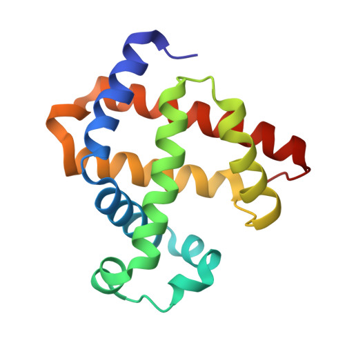
    4TYX

  • PubMed Abstract: 

    Cytochrome c Oxidase (CcO) is known to catalyze the reduction of O2 to H2O efficiently with a much lower overpotential than most other O2 reduction catalysts. However, methods by which the enzyme fine-tunes the reduction potential (E°) of its active site and the corresponding influence on the O2 reduction activity are not well understood. In this work, we report systematic tuning of the heme E° in a functional model of CcO in myoglobin containing three histidines and one tyrosine in the distal pocket of heme. By removing hydrogen-bonding interactions between Ser92 and the proximal His ligand and a heme propionate, and increasing hydrophobicity of the heme pocket through Ser92Ala mutation, we have increased the heme E° from 95 ± 2 to 123 ± 3 mV. Additionally, replacing the native heme b in the CcO mimic with heme a analogs, diacetyl, monoformyl, and diformyl hemes, that posses electron-withdrawing groups, resulted in higher E° values of 175 ± 5, 210 ± 6, and 320 ± 10 mV, respectively. Furthermore, O2 consumption studies on these CcO mimics revealed a strong enhancement in O2 reduction rates with increasing heme E°. Such methods of tuning the heme E° through a combination of secondary sphere mutations and heme substitutions can be applied to tune E° of other heme proteins, allowing for comprehensive investigations of the relationship between E° and enzymatic activity.


  • Organizational Affiliation

    Department of Chemistry, University of Illinois, Urbana-Champaign , Urbana, Illinois 61801, United States.


Macromolecules
Find similar proteins by:  (by identity cutoff)  |  3D Structure
Entity ID: 1
MoleculeChains Sequence LengthOrganismDetailsImage
Myoglobin153Physeter catodonMutation(s): 4 
Gene Names: MB
UniProt
Find proteins for P02185 (Physeter macrocephalus)
Explore P02185 
Go to UniProtKB:  P02185
Entity Groups  
Sequence Clusters30% Identity50% Identity70% Identity90% Identity95% Identity100% Identity
UniProt GroupP02185
Sequence Annotations
Expand
  • Reference Sequence
Small Molecules
Ligands 1 Unique
IDChains Name / Formula / InChI Key2D Diagram3D Interactions
HEM
Query on HEM

Download Ideal Coordinates CCD File 
B [auth A]PROTOPORPHYRIN IX CONTAINING FE
C34 H32 Fe N4 O4
KABFMIBPWCXCRK-RGGAHWMASA-L
Experimental Data & Validation

Experimental Data

  • Method: X-RAY DIFFRACTION
  • Resolution: 1.64 Å
  • R-Value Free: 0.198 
  • R-Value Work: 0.156 
  • R-Value Observed: 0.160 
  • Space Group: P 21 21 21
Unit Cell:
Length ( Å )Angle ( ˚ )
a = 39.467α = 90
b = 47.91β = 90
c = 76.854γ = 90
Software Package:
Software NamePurpose
SCALEPACKdata scaling
CBASSdata collection
PDB_EXTRACTdata extraction
PHENIXrefinement

Structure Validation

View Full Validation Report


Ligand Structure Quality Assessment 


Entry History & Funding Information

Deposition Data


Funding OrganizationLocationGrant Number
National Institutes of Health/National Institute of General Medical Sciences (NIH/NIGMS)United StatesGM062211

Revision History  (Full details and data files)

  • Version 1.0: 2014-08-13
    Type: Initial release
  • Version 1.1: 2014-10-29
    Changes: Database references
  • Version 1.2: 2015-02-04
    Changes: Derived calculations
  • Version 1.3: 2017-09-27
    Changes: Advisory, Author supporting evidence, Derived calculations, Other, Source and taxonomy, Structure summary
  • Version 1.4: 2019-12-25
    Changes: Author supporting evidence
  • Version 1.5: 2023-09-27
    Changes: Data collection, Database references, Refinement description