


Crystal structure of a GroEL D83A/R197A double mutant
Yang, D., Fei, X., LaRonde, N.A., Beckett, D., Lund, P.A., Lorimer, G.H.To be published.
Experimental Data Snapshot
wwPDB Validation 3D Report Full Report

Entity ID: 1 | |||||
|---|---|---|---|---|---|
| Molecule | Chains | Sequence Length | Organism | Details | Image |
| 60 kDa chaperonin | 548 | Escherichia coli K-12 | Mutation(s): 2 Gene Names: groL, groEL, mopA, b4143, JW4103 | 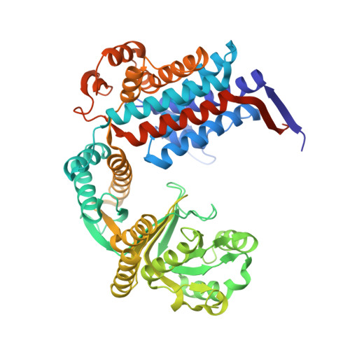 | |
UniProt | |||||
Find proteins for P0A6F5 (Escherichia coli (strain K12)) Explore P0A6F5 Go to UniProtKB: P0A6F5 | |||||
Entity Groups | |||||
| Sequence Clusters | 30% Identity50% Identity70% Identity90% Identity95% Identity100% Identity | ||||
| UniProt Group | P0A6F5 | ||||
Sequence AnnotationsExpand | |||||
| |||||
| Length ( Å ) | Angle ( ˚ ) |
|---|---|
| a = 135.619 | α = 90 |
| b = 259.71 | β = 90 |
| c = 280.848 | γ = 90 |
| Software Name | Purpose |
|---|---|
| PHENIX | refinement |
| iMOSFLM | data reduction |
RCSB PDB (citation) is hosted by
RCSB PDB is a member of the

