4WJG

Structure of T. brucei haptoglobin-hemoglobin receptor binding to human haptoglobin-hemoglobin


Experimental Data Snapshot

  • Method: X-RAY DIFFRACTION
  • Resolution: 3.10 Å
  • R-Value Free: 0.271 
  • R-Value Work: 0.255 
  • R-Value Observed: 0.255 

wwPDB Validation   3D Report Full Report


Ligand Structure Quality Assessment 


This is version 2.0 of the entry. See complete history


Literature

Structural basis for trypanosomal haem acquisition and susceptibility to the host innate immune system.

Stdkilde, K.Torvund-Jensen, M.Moestrup, S.K.Andersen, C.B.

(2014) Nat Commun 5: 5487-5487

  • DOI: https://doi.org/10.1038/ncomms6487
  • Primary Citation of Related Structures:  
    4WJG

  • PubMed Abstract: 

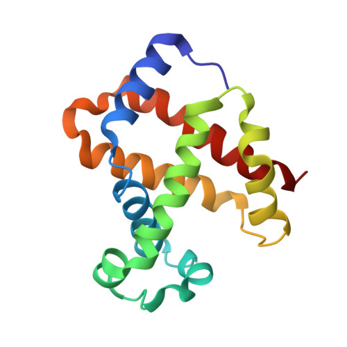
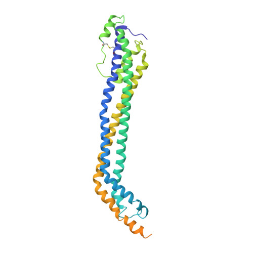
    Sleeping sickness is caused by trypanosome parasites, which infect humans and livestock in Sub-Saharan Africa. Haem is an important growth factor for the parasites and is acquired from the host by receptor-mediated uptake of haptoglobin (Hp)-haemoglobin (Hb) complexes. The parasite Hp-Hb receptor (HpHbR) is also a target for a specialized innate immune defence executed by trypanosome-killing lipoprotein particles containing an Hp-related protein in complex with Hb. Here we report the structure of the multimeric complex between human Hp-Hb and Trypanosoma brucei brucei HpHbR. Two receptors forming kinked three-helical rods with small head regions bind to Hp and the β-subunits of Hb (βHb), with one receptor at each end of the dimeric Hp-Hb complex. The Hb β-subunit haem group directly associates with the receptors, which allows for sensing of haem-containing Hp-Hb. The HpHbR-binding region of Hp is conserved in Hp-related protein, indicating an identical recognition of Hp-Hb and trypanolytic particles by HpHbR in human plasma.


  • Organizational Affiliation

    Department of Biomedicine, Aarhus University, Ole Worms Allé 3, DK-8000 Aarhus C, Denmark.


Macromolecules
Find similar proteins by:  (by identity cutoff)  |  3D Structure
Entity ID: 1
MoleculeChains Sequence LengthOrganismDetailsImage
Hemoglobin subunit alpha
A, F, K, P, U
A, F, K, P, U, Z
141Homo sapiensMutation(s): 0 
UniProt & NIH Common Fund Data Resources
Find proteins for P69905 (Homo sapiens)
Explore P69905 
Go to UniProtKB:  P69905
PHAROS:  P69905
Entity Groups  
Sequence Clusters30% Identity50% Identity70% Identity90% Identity95% Identity100% Identity
UniProt GroupP69905
Sequence Annotations
Expand
  • Reference Sequence
Find similar proteins by:  (by identity cutoff)  |  3D Structure
Entity ID: 2
MoleculeChains Sequence LengthOrganismDetailsImage
Hemoglobin subunit beta146Homo sapiensMutation(s): 0 
UniProt & NIH Common Fund Data Resources
Find proteins for P68871 (Homo sapiens)
Explore P68871 
Go to UniProtKB:  P68871
PHAROS:  P68871
GTEx:  ENSG00000244734 
Entity Groups  
Sequence Clusters30% Identity50% Identity70% Identity90% Identity95% Identity100% Identity
UniProt GroupP68871
Sequence Annotations
Expand
  • Reference Sequence
Find similar proteins by:  (by identity cutoff)  |  3D Structure
Entity ID: 3
MoleculeChains Sequence LengthOrganismDetailsImage
Haptoglobin315Homo sapiensMutation(s): 0 
UniProt & NIH Common Fund Data Resources
Find proteins for P00738 (Homo sapiens)
Explore P00738 
Go to UniProtKB:  P00738
PHAROS:  P00738
GTEx:  ENSG00000257017 
Entity Groups  
Sequence Clusters30% Identity50% Identity70% Identity90% Identity95% Identity100% Identity
UniProt GroupP00738
Sequence Annotations
Expand
  • Reference Sequence
Find similar proteins by:  (by identity cutoff)  |  3D Structure
Entity ID: 4
MoleculeChains Sequence LengthOrganismDetailsImage
Iron-regulated surface determinant protein H146Staphylococcus aureus subsp. aureus N315Mutation(s): 0 
Gene Names: isdHharAsasISA1552
UniProt
Find proteins for Q99TD3 (Staphylococcus aureus (strain N315))
Explore Q99TD3 
Go to UniProtKB:  Q99TD3
Entity Groups  
Sequence Clusters30% Identity50% Identity70% Identity90% Identity95% Identity100% Identity
UniProt GroupQ99TD3
Sequence Annotations
Expand
  • Reference Sequence
Find similar proteins by:  (by identity cutoff)  |  3D Structure
Entity ID: 5
MoleculeChains Sequence LengthOrganismDetailsImage
Haptoglobin-hemoglobin receptor343Trypanosoma brucei bruceiMutation(s): 0 
Gene Names: HpHbR
UniProt
Find proteins for I7BA80 (Trypanosoma brucei brucei)
Explore I7BA80 
Go to UniProtKB:  I7BA80
Entity Groups  
Sequence Clusters30% Identity50% Identity70% Identity90% Identity95% Identity100% Identity
UniProt GroupI7BA80
Sequence Annotations
Expand
  • Reference Sequence
Oligosaccharides

Help

Entity ID: 6
MoleculeChains Length2D Diagram Glycosylation3D Interactions
2-acetamido-2-deoxy-beta-D-glucopyranose-(1-4)-2-acetamido-2-deoxy-beta-D-glucopyranose
EA [auth a],
FA [auth b],
GA [auth c],
HA [auth d],
IA [auth e],
EA [auth a],
FA [auth b],
GA [auth c],
HA [auth d],
IA [auth e],
JA [auth f],
KA [auth g],
LA [auth h],
MA [auth i],
NA [auth j]
2N-Glycosylation
Glycosylation Resources
GlyTouCan:  G42666HT
GlyCosmos:  G42666HT
GlyGen:  G42666HT
Small Molecules
Ligands 3 Unique
IDChains Name / Formula / InChI Key2D Diagram3D Interactions
HEM
Query on HEM

Download Ideal Coordinates CCD File 
CC [auth Z]
EB [auth K]
EC [auth 1]
GB [auth L]
MB [auth P]
CC [auth Z],
EB [auth K],
EC [auth 1],
GB [auth L],
MB [auth P],
OA [auth A],
OB [auth Q],
QA [auth B],
UB [auth U],
WA [auth F],
WB [auth V],
YA [auth G]
PROTOPORPHYRIN IX CONTAINING FE
C34 H32 Fe N4 O4
KABFMIBPWCXCRK-RGGAHWMASA-L
NAG
Query on NAG

Download Ideal Coordinates CCD File 
AB [auth H]
AC [auth Y]
BB [auth H]
BC [auth Y]
CB [auth J]
AB [auth H],
AC [auth Y],
BB [auth H],
BC [auth Y],
CB [auth J],
DB [auth J],
GC [auth 2],
HC [auth 4],
IB [auth M],
IC [auth 4],
JB [auth M],
KB [auth O],
LB [auth O],
QB [auth R],
RB [auth R],
SA [auth C],
SB [auth T],
TA [auth C],
TB [auth T],
UA [auth E],
VA [auth E],
YB [auth W],
ZB [auth W]
2-acetamido-2-deoxy-beta-D-glucopyranose
C8 H15 N O6
OVRNDRQMDRJTHS-FMDGEEDCSA-N
OXY
Query on OXY

Download Ideal Coordinates CCD File 
DC [auth Z]
FB [auth K]
FC [auth 1]
HB [auth L]
NB [auth P]
DC [auth Z],
FB [auth K],
FC [auth 1],
HB [auth L],
NB [auth P],
PA [auth A],
PB [auth Q],
RA [auth B],
VB [auth U],
XA [auth F],
XB [auth V],
ZA [auth G]
OXYGEN MOLECULE
O2
MYMOFIZGZYHOMD-UHFFFAOYSA-N
Experimental Data & Validation

Experimental Data

  • Method: X-RAY DIFFRACTION
  • Resolution: 3.10 Å
  • R-Value Free: 0.271 
  • R-Value Work: 0.255 
  • R-Value Observed: 0.255 
  • Space Group: P 1 21 1
Unit Cell:
Length ( Å )Angle ( ˚ )
a = 143.23α = 90
b = 140.95β = 98.54
c = 267.18γ = 90
Software Package:
Software NamePurpose
PHENIXrefinement
XDSdata reduction
XSCALEdata scaling
PHASERphasing

Structure Validation

View Full Validation Report


Ligand Structure Quality Assessment 


Entry History & Funding Information

Deposition Data


Funding OrganizationLocationGrant Number
The Lundbeck FoundationDenmark--
The Novo Nordisk FoundationDenmark--
The European Research CouncilDenmark--
The Danish Council for Independent ResearchDenmark--

Revision History  (Full details and data files)

  • Version 1.0: 2014-11-26
    Type: Initial release
  • Version 1.1: 2014-12-03
    Changes: Database references
  • Version 1.2: 2018-02-28
    Changes: Source and taxonomy
  • Version 2.0: 2020-07-29
    Type: Remediation
    Reason: Carbohydrate remediation
    Changes: Advisory, Atomic model, Data collection, Derived calculations, Source and taxonomy, Structure summary