4XR8

Crystal structure of the HPV16 E6/E6AP/p53 ternary complex at 2.25 A resolution


Experimental Data Snapshot

  • Method: X-RAY DIFFRACTION
  • Resolution: 2.25 Å
  • R-Value Free: 0.246 
  • R-Value Work: 0.194 
  • R-Value Observed: 0.196 

wwPDB Validation   3D Report Full Report


This is version 2.1 of the entry. See complete history


Literature

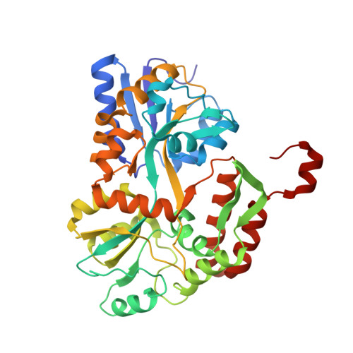
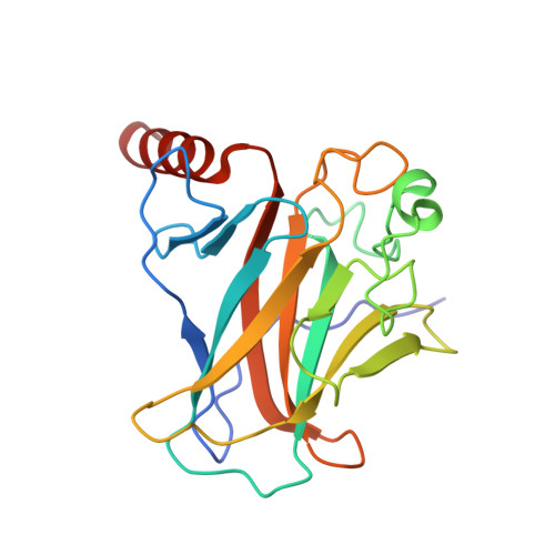
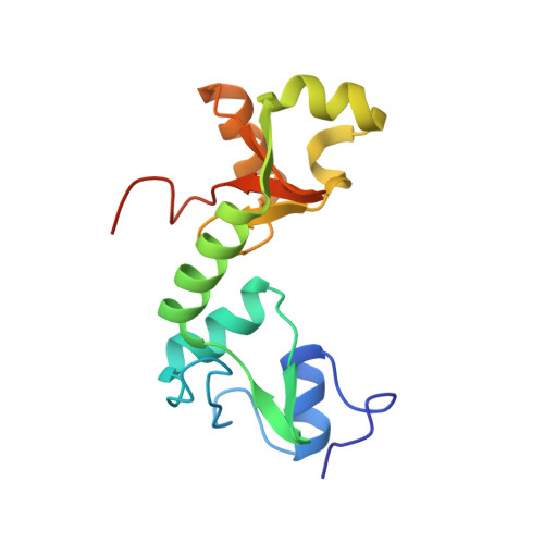
Structure of the E6/E6AP/p53 complex required for HPV-mediated degradation of p53.

Martinez-Zapien, D.Ruiz, F.X.Poirson, J.Mitschler, A.Ramirez, J.Forster, A.Cousido-Siah, A.Masson, M.Pol, S.V.Podjarny, A.Trave, G.Zanier, K.

(2016) Nature 529: 541-545

  • DOI: https://doi.org/10.1038/nature16481
  • Primary Citation of Related Structures:  
    4XR8

  • PubMed Abstract: 

    The p53 pro-apoptotic tumour suppressor is mutated or functionally altered in most cancers. In epithelial tumours induced by 'high-risk' mucosal human papilloma viruses, including human cervical carcinoma and a growing number of head-and-neck cancers, p53 is degraded by the viral oncoprotein E6 (ref. 2). In this process, E6 binds to a short leucine (L)-rich LxxLL consensus sequence within the cellular ubiquitin ligase E6AP. Subsequently, the E6/E6AP heterodimer recruits and degrades p53 (ref. 4). Neither E6 nor E6AP are separately able to recruit p53 (refs 3, 5), and the precise mode of assembly of E6, E6AP and p53 is unknown. Here we solve the crystal structure of a ternary complex comprising full-length human papilloma virus type 16 (HPV-16) E6, the LxxLL motif of E6AP and the core domain of p53. The LxxLL motif of E6AP renders the conformation of E6 competent for interaction with p53 by structuring a p53-binding cleft on E6. Mutagenesis of critical positions at the E6-p53 interface disrupts p53 degradation. The E6-binding site of p53 is distal from previously described DNA- and protein-binding surfaces of the core domain. This suggests that, in principle, E6 may avoid competition with cellular factors by targeting both free and bound p53 molecules. The E6/E6AP/p53 complex represents a prototype of viral hijacking of both the ubiquitin-mediated protein degradation pathway and the p53 tumour suppressor pathway. The present structure provides a framework for the design of inhibitory therapeutic strategies against oncogenesis mediated by human papilloma virus.


  • Organizational Affiliation

    Equipe labellisée Ligue, Biotechnologie et signalisation cellulaire UMR 7242, Ecole Superieure de Biotechnologie de Strasbourg, Boulevard Sébastien Brant, BP 10413, F-67412 Illkirch, France.


Macromolecules
Find similar proteins by:  (by identity cutoff)  |  3D Structure
Entity ID: 1
MoleculeChains Sequence LengthOrganismDetailsImage
Maltose-binding periplasmic protein, ubiquitin ligase E6AP
A, B
383Escherichia coli K-12Homo sapiens
This entity is chimeric
Mutation(s): 6 
Gene Names: malEb4034JW3994
UniProt & NIH Common Fund Data Resources
Find proteins for P0AEX9 (Escherichia coli (strain K12))
Explore P0AEX9 
Go to UniProtKB:  P0AEX9
Find proteins for Q05086 (Homo sapiens)
Explore Q05086 
Go to UniProtKB:  Q05086
PHAROS:  Q05086
GTEx:  ENSG00000114062 
Entity Groups  
Sequence Clusters30% Identity50% Identity70% Identity90% Identity95% Identity100% Identity
UniProt GroupsQ05086P0AEX9
Sequence Annotations
Expand
  • Reference Sequence
Find similar proteins by:  (by identity cutoff)  |  3D Structure
Entity ID: 2
MoleculeChains Sequence LengthOrganismDetailsImage
Cellular tumor antigen p53
C, D
199Homo sapiensMutation(s): 0 
Gene Names: TP53P53
UniProt & NIH Common Fund Data Resources
Find proteins for P04637 (Homo sapiens)
Explore P04637 
Go to UniProtKB:  P04637
PHAROS:  P04637
GTEx:  ENSG00000141510 
Entity Groups  
Sequence Clusters30% Identity50% Identity70% Identity90% Identity95% Identity100% Identity
UniProt GroupP04637
Sequence Annotations
Expand
  • Reference Sequence
Find similar proteins by:  (by identity cutoff)  |  3D Structure
Entity ID: 3
MoleculeChains Sequence LengthOrganismDetailsImage
Protein E6E [auth F],
F [auth H]
151Human papillomavirus 16Mutation(s): 4 
Gene Names: E6
UniProt
Find proteins for P03126 (Human papillomavirus type 16)
Explore P03126 
Go to UniProtKB:  P03126
Entity Groups  
Sequence Clusters30% Identity50% Identity70% Identity90% Identity95% Identity100% Identity
UniProt GroupP03126
Sequence Annotations
Expand
  • Reference Sequence
Oligosaccharides

Help

Entity ID: 4
MoleculeChains Length2D Diagram Glycosylation3D Interactions
alpha-D-glucopyranose-(1-4)-alpha-D-glucopyranoseG [auth E],
H [auth G]
2N/A
Glycosylation Resources
GlyTouCan:  G07411ON
GlyCosmos:  G07411ON
Small Molecules
Ligands 3 Unique
IDChains Name / Formula / InChI Key2D Diagram3D Interactions
PEG
Query on PEG

Download Ideal Coordinates CCD File 
I [auth A],
J [auth B],
M [auth D],
Q [auth F]
DI(HYDROXYETHYL)ETHER
C4 H10 O3
MTHSVFCYNBDYFN-UHFFFAOYSA-N
ZN
Query on ZN

Download Ideal Coordinates CCD File 
K [auth C]
L [auth D]
N [auth F]
O [auth F]
R [auth H]
K [auth C],
L [auth D],
N [auth F],
O [auth F],
R [auth H],
S [auth H]
ZINC ION
Zn
PTFCDOFLOPIGGS-UHFFFAOYSA-N
EDO
Query on EDO

Download Ideal Coordinates CCD File 
P [auth F]1,2-ETHANEDIOL
C2 H6 O2
LYCAIKOWRPUZTN-UHFFFAOYSA-N
Biologically Interesting Molecules (External Reference) 1 Unique
Experimental Data & Validation

Experimental Data

  • Method: X-RAY DIFFRACTION
  • Resolution: 2.25 Å
  • R-Value Free: 0.246 
  • R-Value Work: 0.194 
  • R-Value Observed: 0.196 
  • Space Group: P 1 21 1
Unit Cell:
Length ( Å )Angle ( ˚ )
a = 77.842α = 90
b = 128.939β = 92.33
c = 81.56γ = 90
Software Package:
Software NamePurpose
HKL-2000data reduction
HKL-2000data scaling
PHASERphasing
PHENIXrefinement

Structure Validation

View Full Validation Report


Entry History 

Deposition Data

Revision History  (Full details and data files)

  • Version 1.0: 2016-02-03
    Type: Initial release
  • Version 2.0: 2020-07-29
    Type: Remediation
    Reason: Carbohydrate remediation
    Changes: Atomic model, Data collection, Derived calculations, Non-polymer description, Structure summary
  • Version 2.1: 2024-01-10
    Changes: Data collection, Database references, Refinement description, Structure summary