4XVU

Structure of Get3 bound to the transmembrane domain of Nyv1


Experimental Data Snapshot

  • Method: X-RAY DIFFRACTION
  • Resolution: 2.35 Å
  • R-Value Free: 0.234 
  • R-Value Work: 0.197 
  • R-Value Observed: 0.199 

wwPDB Validation   3D Report Full Report


Ligand Structure Quality Assessment 


This is version 1.3 of the entry. See complete history


Literature

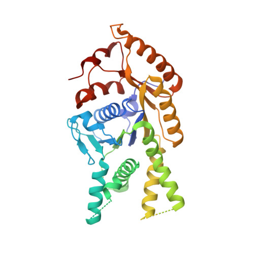
Protein targeting. Structure of the Get3 targeting factor in complex with its membrane protein cargo.

Mateja, A.Paduch, M.Chang, H.Y.Szydlowska, A.Kossiakoff, A.A.Hegde, R.S.Keenan, R.J.

(2015) Science 347: 1152-1155

  • DOI: https://doi.org/10.1126/science.1261671
  • Primary Citation of Related Structures:  
    4XTR, 4XVU, 4XWO

  • PubMed Abstract: 

    Tail-anchored (TA) proteins are a physiologically important class of membrane proteins targeted to the endoplasmic reticulum by the conserved guided-entry of TA proteins (GET) pathway. During transit, their hydrophobic transmembrane domains (TMDs) are chaperoned by the cytosolic targeting factor Get3, but the molecular nature of the functional Get3-TA protein targeting complex remains unknown. We reconstituted the physiologic assembly pathway for a functional targeting complex and showed that it comprises a TA protein bound to a Get3 homodimer. Crystal structures of Get3 bound to different TA proteins showed an α-helical TMD occupying a hydrophobic groove that spans the Get3 homodimer. Our data elucidate the mechanism of TA protein recognition and shielding by Get3 and suggest general principles of hydrophobic domain chaperoning by cellular targeting factors.


  • Organizational Affiliation

    Department of Biochemistry and Molecular Biology, The University of Chicago, 929 East 57th Street, Chicago, IL 60637, USA.


Macromolecules
Find similar proteins by:  (by identity cutoff)  |  3D Structure
Entity ID: 1
MoleculeChains Sequence LengthOrganismDetailsImage
ATPase GET3
A, B, G, H
354Saccharomyces cerevisiae S288CMutation(s): 1 
Gene Names: GET3ARR4YDL100CD2371
EC: 3.6
UniProt
Find proteins for Q12154 (Saccharomyces cerevisiae (strain ATCC 204508 / S288c))
Explore Q12154 
Go to UniProtKB:  Q12154
Entity Groups  
Sequence Clusters30% Identity50% Identity70% Identity90% Identity95% Identity100% Identity
UniProt GroupQ12154
Sequence Annotations
Expand
  • Reference Sequence
Find similar proteins by:  (by identity cutoff)  |  3D Structure
Entity ID: 2
MoleculeChains Sequence LengthOrganismDetailsImage
Antibody heavy chain
C, E, I, K
230Homo sapienssynthetic construct
This entity is chimeric
Mutation(s): 0 
Entity Groups  
Sequence Clusters30% Identity50% Identity70% Identity90% Identity95% Identity100% Identity
Sequence Annotations
Expand
  • Reference Sequence
Find similar proteins by:  (by identity cutoff)  |  3D Structure
Entity ID: 3
MoleculeChains Sequence LengthOrganismDetailsImage
Antibody light chain
D, F, J, L
217Homo sapienssynthetic construct
This entity is chimeric
Mutation(s): 0 
Entity Groups  
Sequence Clusters30% Identity50% Identity70% Identity90% Identity95% Identity100% Identity
Sequence Annotations
Expand
  • Reference Sequence
Find similar proteins by:  (by identity cutoff)  |  3D Structure
Entity ID: 4
MoleculeChains Sequence LengthOrganismDetailsImage
Nyv1 TMDM [auth g],
N [auth a]
37Saccharomyces cerevisiaeMutation(s): 0 
Sequence Annotations
Expand
  • Reference Sequence
Small Molecules
Ligands 3 Unique
IDChains Name / Formula / InChI Key2D Diagram3D Interactions
ATP
Query on ATP

Download Ideal Coordinates CCD File 
O [auth A],
R [auth B],
T [auth G],
W [auth H]
ADENOSINE-5'-TRIPHOSPHATE
C10 H16 N5 O13 P3
ZKHQWZAMYRWXGA-KQYNXXCUSA-N
ZN
Query on ZN

Download Ideal Coordinates CCD File 
Q [auth A],
V [auth G]
ZINC ION
Zn
PTFCDOFLOPIGGS-UHFFFAOYSA-N
MG
Query on MG

Download Ideal Coordinates CCD File 
P [auth A],
S [auth B],
U [auth G],
X [auth H]
MAGNESIUM ION
Mg
JLVVSXFLKOJNIY-UHFFFAOYSA-N
Experimental Data & Validation

Experimental Data

  • Method: X-RAY DIFFRACTION
  • Resolution: 2.35 Å
  • R-Value Free: 0.234 
  • R-Value Work: 0.197 
  • R-Value Observed: 0.199 
  • Space Group: P 1
Unit Cell:
Length ( Å )Angle ( ˚ )
a = 79.44α = 63.05
b = 109.23β = 77.74
c = 111.35γ = 70.17
Software Package:
Software NamePurpose
PHENIXrefinement
xia2data reduction
PHASERphasing
Cootmodel building

Structure Validation

View Full Validation Report


Ligand Structure Quality Assessment 


Entry History & Funding Information

Deposition Data


Funding OrganizationLocationGrant Number
National Institutes of Health/National Institute of General Medical Sciences (NIH/NIGMS)United StatesR01 GM086487
National Institutes of Health/National Institute of General Medical Sciences (NIH/NIGMS)United StatesU01 GM094588
National Institutes of Health/National Institute of General Medical Sciences (NIH/NIGMS)United StatesU54 GM087519

Revision History  (Full details and data files)

  • Version 1.0: 2015-03-18
    Type: Initial release
  • Version 1.1: 2017-09-20
    Changes: Author supporting evidence, Derived calculations, Source and taxonomy
  • Version 1.2: 2019-12-25
    Changes: Author supporting evidence
  • Version 1.3: 2023-09-27
    Changes: Data collection, Database references, Derived calculations, Refinement description