4YFK

Escherichia coli RNA polymerase in complex with squaramide compound 8.


Experimental Data Snapshot

  • Method: X-RAY DIFFRACTION
  • Resolution: 3.57 Å
  • R-Value Free: 0.252 
  • R-Value Work: 0.217 
  • R-Value Observed: 0.217 

wwPDB Validation   3D Report Full Report


Ligand Structure Quality Assessment 


This is version 1.5 of the entry. See complete history


Literature

X-ray Crystal Structures of Escherichia coli RNA Polymerase with Switch Region Binding Inhibitors Enable Rational Design of Squaramides with an Improved Fraction Unbound to Human Plasma Protein.

Molodtsov, V.Fleming, P.R.Eyermann, C.J.Ferguson, A.D.Foulk, M.A.McKinney, D.C.Masse, C.E.Buurman, E.T.Murakami, K.S.

(2015) J Med Chem 58: 3156-3171

  • DOI: https://doi.org/10.1021/acs.jmedchem.5b00050
  • Primary Citation of Related Structures:  
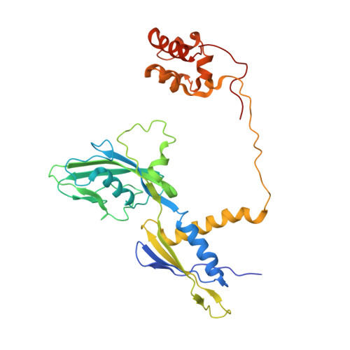
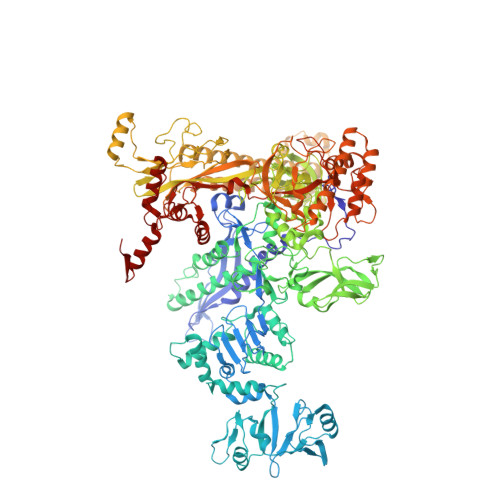
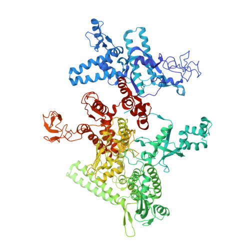
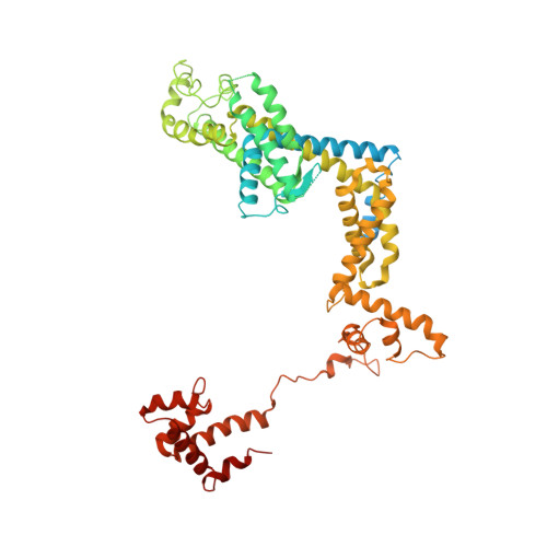
    4YFK, 4YFN, 4YFX

  • PubMed Abstract: 

    Squaramides constitute a novel class of RNA polymerase inhibitors of which genetic evidence and computational modeling previously have suggested an inhibitory mechanism mediated by binding to the RNA polymerase switch region. An iterative chemistry program increased the fraction unbound to human plasma protein from below minimum detection levels, i.e., <1% to 4-6%, while retaining biochemical potency. Since in vitro antimicrobial activity against an efflux-negative strain of Haemophilus influenzae was 4- to 8-fold higher, the combined improvement was at least 20- to 60-fold. Cocrystal structures of Escherichia coli RNA polymerase with two key squaramides showed displacement of the switch 2, predicted to interfere with the conformational change of the clamp domain and/or with binding of template DNA, a mechanism akin to that of natural product myxopyronin. Furthermore, the structures confirmed the chemical features required for biochemical potency. The terminal isoxazole and benzyl rings bind into distinct relatively narrow, hydrophobic pockets, and both are required for biochemical potency. In contrast, the linker composed of squarate and piperidine accesses different conformations in their respective cocrystal structures with RNA polymerase, reflecting its main role of proper orientation of the aforementioned terminal rings. These observations further explain the tolerance of hydrophilic substitutions in the linker region that was exploited to improve the fraction unbound to human plasma protein while retaining biochemical potency.


  • Organizational Affiliation

    †Department of Biochemistry and Molecular Biology, The Center for RNA Molecular Biology, The Pennsylvania State University, University Park, Pennsylvania 16802, United States.


Macromolecules
Find similar proteins by:  (by identity cutoff)  |  3D Structure
Entity ID: 1
MoleculeChains Sequence LengthOrganismDetailsImage
DNA-directed RNA polymerase subunit alpha
A, B, G, H
329Escherichia coli O139:H28 str. E24377AMutation(s): 0 
Gene Names: rpoAEcE24377A_3778
EC: 2.7.7.6
UniProt
Find proteins for A7ZSI4 (Escherichia coli O139:H28 (strain E24377A / ETEC))
Explore A7ZSI4 
Go to UniProtKB:  A7ZSI4
Entity Groups  
Sequence Clusters30% Identity50% Identity70% Identity90% Identity95% Identity100% Identity
UniProt GroupA7ZSI4
Sequence Annotations
Expand
  • Reference Sequence
Find similar proteins by:  (by identity cutoff)  |  3D Structure
Entity ID: 2
MoleculeChains Sequence LengthOrganismDetailsImage
DNA-directed RNA polymerase subunit beta
C, I
1,342Escherichia coli O139:H28 str. E24377AMutation(s): 0 
Gene Names: rpoBEcE24377A_4528
EC: 2.7.7.6
UniProt
Find proteins for A7ZUK1 (Escherichia coli O139:H28 (strain E24377A / ETEC))
Explore A7ZUK1 
Go to UniProtKB:  A7ZUK1
Entity Groups  
Sequence Clusters30% Identity50% Identity70% Identity90% Identity95% Identity100% Identity
UniProt GroupA7ZUK1
Sequence Annotations
Expand
  • Reference Sequence
Find similar proteins by:  (by identity cutoff)  |  3D Structure
Entity ID: 3
MoleculeChains Sequence LengthOrganismDetailsImage
DNA-directed RNA polymerase subunit beta'
D, J
1,407Escherichia coli O139:H28 str. E24377AMutation(s): 0 
Gene Names: rpoCEcE24377A_4529
EC: 2.7.7.6
UniProt
Find proteins for A7ZUK2 (Escherichia coli O139:H28 (strain E24377A / ETEC))
Explore A7ZUK2 
Go to UniProtKB:  A7ZUK2
Entity Groups  
Sequence Clusters30% Identity50% Identity70% Identity90% Identity95% Identity100% Identity
UniProt GroupA7ZUK2
Sequence Annotations
Expand
  • Reference Sequence
Find similar proteins by:  (by identity cutoff)  |  3D Structure
Entity ID: 4
MoleculeChains Sequence LengthOrganismDetailsImage
DNA-directed RNA polymerase subunit omega
E, K
91Escherichia coli O139:H28 str. E24377AMutation(s): 0 
Gene Names: rpoZEcE24377A_4152
EC: 2.7.7.6
UniProt
Find proteins for A7ZTK1 (Escherichia coli O139:H28 (strain E24377A / ETEC))
Explore A7ZTK1 
Go to UniProtKB:  A7ZTK1
Entity Groups  
Sequence Clusters30% Identity50% Identity70% Identity90% Identity95% Identity100% Identity
UniProt GroupA7ZTK1
Sequence Annotations
Expand
  • Reference Sequence
Find similar proteins by:  (by identity cutoff)  |  3D Structure
Entity ID: 5
MoleculeChains Sequence LengthOrganismDetailsImage
RNA polymerase sigma factor RpoD
F, L
613Escherichia coli K-12Mutation(s): 0 
Gene Names: rpoDaltb3067JW3039
UniProt
Find proteins for P00579 (Escherichia coli (strain K12))
Explore P00579 
Go to UniProtKB:  P00579
Entity Groups  
Sequence Clusters30% Identity50% Identity70% Identity90% Identity95% Identity100% Identity
UniProt GroupP00579
Sequence Annotations
Expand
  • Reference Sequence
Small Molecules
Ligands 3 Unique
IDChains Name / Formula / InChI Key2D Diagram3D Interactions
4C6
Query on 4C6

Download Ideal Coordinates CCD File 
Q [auth D],
V [auth J]
3,5-dimethyl-N-{2-[4-(4-methylbenzyl)piperidin-1-yl]-3,4-dioxocyclobut-1-en-1-yl}-1,2-oxazole-4-sulfonamide
C22 H25 N3 O5 S
DJQHTHRCUXNHLQ-UHFFFAOYSA-N
ZN
Query on ZN

Download Ideal Coordinates CCD File 
O [auth D],
P [auth D],
T [auth J],
U [auth J]
ZINC ION
Zn
PTFCDOFLOPIGGS-UHFFFAOYSA-N
MG
Query on MG

Download Ideal Coordinates CCD File 
M [auth C],
N [auth D],
R [auth I],
S [auth J]
MAGNESIUM ION
Mg
JLVVSXFLKOJNIY-UHFFFAOYSA-N
Experimental Data & Validation

Experimental Data

  • Method: X-RAY DIFFRACTION
  • Resolution: 3.57 Å
  • R-Value Free: 0.252 
  • R-Value Work: 0.217 
  • R-Value Observed: 0.217 
  • Space Group: P 21 21 21
Unit Cell:
Length ( Å )Angle ( ˚ )
a = 186.105α = 90
b = 206.438β = 90
c = 307.993γ = 90
Software Package:
Software NamePurpose
PHENIXrefinement
HKL-2000data scaling
PHASERphasing
Cootmodel building

Structure Validation

View Full Validation Report


Ligand Structure Quality Assessment 


Entry History & Funding Information

Deposition Data


Funding OrganizationLocationGrant Number
National Institutes of Health/National Institute of General Medical Sciences (NIH/NIGMS)United StatesGM087350

Revision History  (Full details and data files)

  • Version 1.0: 2015-03-11
    Type: Initial release
  • Version 1.1: 2015-04-08
    Changes: Database references
  • Version 1.2: 2015-04-22
    Changes: Database references
  • Version 1.3: 2017-09-20
    Changes: Author supporting evidence, Database references, Derived calculations, Refinement description, Source and taxonomy
  • Version 1.4: 2019-12-25
    Changes: Author supporting evidence
  • Version 1.5: 2023-09-27
    Changes: Data collection, Database references, Derived calculations, Refinement description