4ZFP

A new crystal structure for the adduct formed in the reaction between AuSac2, a cytotoxic homoleptic gold(I) compound with the saccharinate ligand, and the model protein hen egg white lysozyme


Experimental Data Snapshot

  • Method: X-RAY DIFFRACTION
  • Resolution: 1.96 Å
  • R-Value Free: 0.235 
  • R-Value Work: 0.170 
  • R-Value Observed: 0.173 

wwPDB Validation   3D Report Full Report


This is version 1.2 of the entry. See complete history


Literature

Structural evidences for a secondary gold binding site in the hydrophobic box of lysozyme.

Ferraro, G.Massai, L.Messori, L.Cinellu, M.A.Merlino, A.

(2015) Biometals 28: 745-754

  • DOI: https://doi.org/10.1007/s10534-015-9863-7
  • Primary Citation of Related Structures:  
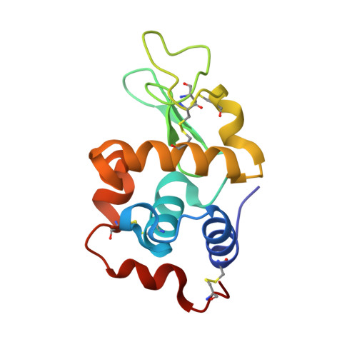
    4ZFP

  • PubMed Abstract: 

    A new crystal structure is reported here for the adduct formed in the reaction between NH4 [Au(Sac)2], AuSac2, a cytotoxic homoleptic gold(I) complex with the saccharinate ligand, and the model protein hen egg white lysozyme. To produce this adduct, AuSac2 breaks down and releases both saccharinate ligands. The resulting Au(I) ions bind the protein to ND1 and NE2 atoms of His15 but also to SD atom of the zero-solvent accessible Met105 side chain, which is located in the protein hydrophobic box. The unexpected existence of this secondary gold(I) binding site is confirmed by spectroscopic and spectrometric measurements in solution.


  • Organizational Affiliation

    Department of Chemical Sciences, University of Naples Federico II, Via Cintia, 80126, Naples, Italy.


Macromolecules
Find similar proteins by:  (by identity cutoff)  |  3D Structure
Entity ID: 1
MoleculeChains Sequence LengthOrganismDetailsImage
Lysozyme C129Gallus gallusMutation(s): 0 
EC: 3.2.1.17
UniProt
Find proteins for P00698 (Gallus gallus)
Explore P00698 
Go to UniProtKB:  P00698
Entity Groups  
Sequence Clusters30% Identity50% Identity70% Identity90% Identity95% Identity100% Identity
UniProt GroupP00698
Sequence Annotations
Expand
  • Reference Sequence
Small Molecules
Ligands 3 Unique
IDChains Name / Formula / InChI Key2D Diagram3D Interactions
AU
Query on AU

Download Ideal Coordinates CCD File 
I [auth A],
J [auth A],
K [auth A]
GOLD ION
Au
ZBKIUFWVEIBQRT-UHFFFAOYSA-N
EDO
Query on EDO

Download Ideal Coordinates CCD File 
B [auth A]1,2-ETHANEDIOL
C2 H6 O2
LYCAIKOWRPUZTN-UHFFFAOYSA-N
NO3
Query on NO3

Download Ideal Coordinates CCD File 
C [auth A]
D [auth A]
E [auth A]
F [auth A]
G [auth A]
C [auth A],
D [auth A],
E [auth A],
F [auth A],
G [auth A],
H [auth A]
NITRATE ION
N O3
NHNBFGGVMKEFGY-UHFFFAOYSA-N
Experimental Data & Validation

Experimental Data

  • Method: X-RAY DIFFRACTION
  • Resolution: 1.96 Å
  • R-Value Free: 0.235 
  • R-Value Work: 0.170 
  • R-Value Observed: 0.173 
  • Space Group: P 43 21 2
Unit Cell:
Length ( Å )Angle ( ˚ )
a = 77.584α = 90
b = 77.584β = 90
c = 37.111γ = 90
Software Package:
Software NamePurpose
REFMACrefinement
HKL-2000data reduction
HKL-2000data scaling
REFMACphasing

Structure Validation

View Full Validation Report


Entry History 

Deposition Data

  • Released Date: 2015-06-24 
  • Deposition Author(s): Merlino, A.

Revision History  (Full details and data files)

  • Version 1.0: 2015-06-24
    Type: Initial release
  • Version 1.1: 2015-07-08
    Changes: Database references
  • Version 1.2: 2024-01-10
    Changes: Data collection, Database references, Derived calculations, Refinement description