4ZNC

Fc fragment of human IgG in complex with the C domain of staphylococcal protein A mutant - Q9W


Experimental Data Snapshot

  • Method: X-RAY DIFFRACTION
  • Resolution: 2.28 Å
  • R-Value Free: 0.242 
  • R-Value Work: 0.196 
  • R-Value Observed: 0.198 

wwPDB Validation   3D Report Full Report


This is version 1.3 of the entry. See complete history


Literature

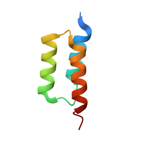
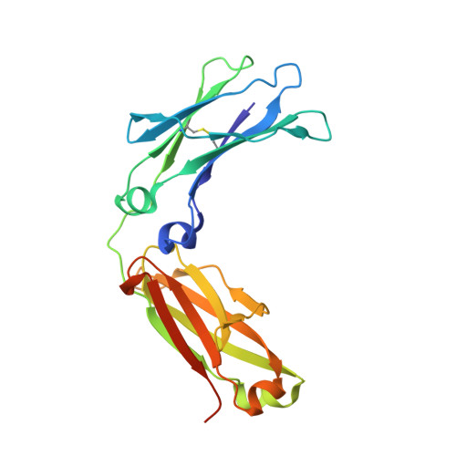
Suppression of conformational heterogeneity at a protein-protein interface.

Deis, L.N.Wu, Q.Wang, Y.Qi, Y.Daniels, K.G.Zhou, P.Oas, T.G.

(2015) Proc Natl Acad Sci U S A 112: 9028-9033

  • DOI: https://doi.org/10.1073/pnas.1424724112
  • Primary Citation of Related Structures:  
    4WWI, 4ZMD, 4ZNC

  • PubMed Abstract: 

    Staphylococcal protein A (SpA) is an important virulence factor from Staphylococcus aureus responsible for the bacterium's evasion of the host immune system. SpA includes five small three-helix-bundle domains that can each bind with high affinity to many host proteins such as antibodies. The interaction between a SpA domain and the Fc fragment of IgG was partially elucidated previously in the crystal structure 1FC2. Although informative, the previous structure was not properly folded and left many substantial questions unanswered, such as a detailed description of the tertiary structure of SpA domains in complex with Fc and the structural changes that take place upon binding. Here we report the 2.3-Å structure of a fully folded SpA domain in complex with Fc. Our structure indicates that there are extensive structural rearrangements necessary for binding Fc, including a general reduction in SpA conformational heterogeneity, freezing out of polyrotameric interfacial residues, and displacement of a SpA side chain by an Fc side chain in a molecular-recognition pocket. Such a loss of conformational heterogeneity upon formation of the protein-protein interface may occur when SpA binds its multiple binding partners. Suppression of conformational heterogeneity may be an important structural paradigm in functionally plastic proteins.


  • Organizational Affiliation

    Department of Biochemistry, Duke University, Durham, NC 27710.


Macromolecules
Find similar proteins by:  (by identity cutoff)  |  3D Structure
Entity ID: 1
MoleculeChains Sequence LengthOrganismDetailsImage
Immunoglobulin G-binding protein A
A, B, C
58Staphylococcus aureusMutation(s): 1 
Gene Names: spa
UniProt
Find proteins for P38507 (Staphylococcus aureus)
Explore P38507 
Go to UniProtKB:  P38507
Entity Groups  
Sequence Clusters30% Identity50% Identity70% Identity90% Identity95% Identity100% Identity
UniProt GroupP38507
Sequence Annotations
Expand
  • Reference Sequence
Find similar proteins by:  (by identity cutoff)  |  3D Structure
Entity ID: 2
MoleculeChains Sequence LengthOrganismDetailsImage
Ig gamma-3 chain C region
D, E, F
220Homo sapiensMutation(s): 0 
Gene Names: IGHG3
UniProt & NIH Common Fund Data Resources
Find proteins for P01860 (Homo sapiens)
Explore P01860 
Go to UniProtKB:  P01860
PHAROS:  P01860
Entity Groups  
Sequence Clusters30% Identity50% Identity70% Identity90% Identity95% Identity100% Identity
UniProt GroupP01860
Sequence Annotations
Expand
  • Reference Sequence
Experimental Data & Validation

Experimental Data

  • Method: X-RAY DIFFRACTION
  • Resolution: 2.28 Å
  • R-Value Free: 0.242 
  • R-Value Work: 0.196 
  • R-Value Observed: 0.198 
  • Space Group: C 1 2 1
Unit Cell:
Length ( Å )Angle ( ˚ )
a = 137.951α = 90
b = 87.21β = 91.06
c = 103.248γ = 90
Software Package:
Software NamePurpose
PHENIXrefinement
HKL-2000data reduction
HKL-2000data scaling
PHASERphasing

Structure Validation

View Full Validation Report


Entry History 

Deposition Data

Revision History  (Full details and data files)

  • Version 1.0: 2015-07-15
    Type: Initial release
  • Version 1.1: 2015-07-22
    Changes: Derived calculations
  • Version 1.2: 2015-07-29
    Changes: Database references
  • Version 1.3: 2023-09-27
    Changes: Data collection, Database references, Derived calculations, Refinement description, Source and taxonomy