5BW7

Crystal structure of nonfucosylated Fc Y296W mutant complexed with bis-glycosylated soluble form of Fc gamma receptor IIIa


Experimental Data Snapshot

  • Method: X-RAY DIFFRACTION
  • Resolution: 3.00 Å
  • R-Value Free: 0.250 
  • R-Value Work: 0.204 
  • R-Value Observed: 0.206 

wwPDB Validation   3D Report Full Report


This is version 2.2 of the entry. See complete history


Literature

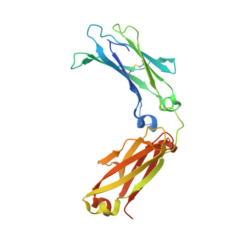
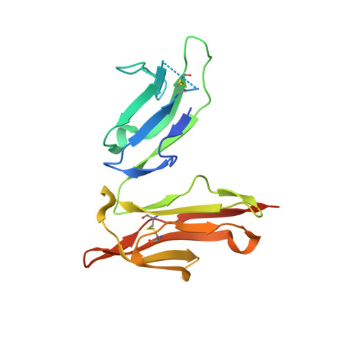
Importance of the Side Chain at Position 296 of Antibody Fc in Interactions with Fc gamma RIIIa and Other Fc gamma Receptors

Isoda, Y.Yagi, H.Satoh, T.Shibata-Koyama, M.Masuda, K.Satoh, M.Kato, K.Iida, S.

(2015) PLoS One 10: e0140120-e0140120

  • DOI: https://doi.org/10.1371/journal.pone.0140120
  • Primary Citation of Related Structures:  
    5BW7

  • PubMed Abstract: 

    Antibody-dependent cellular cytotoxicity (ADCC) is an important effector function determining the clinical efficacy of therapeutic antibodies. Core fucose removal from N-glycans on the Fc portion of immunoglobulin G (IgG) improves the binding affinity for Fcγ receptor IIIa (FcγRIIIa) and dramatically enhances ADCC. Our previous structural analyses revealed that Tyr-296 of IgG1-Fc plays a critical role in the interaction with FcγRIIIa, particularly in the enhanced FcγRIIIa binding of nonfucosylated IgG1. However, the importance of the Tyr-296 residue in the antibody in the interaction with various Fcγ receptors has not yet been elucidated. To further clarify the biological importance of this residue, we established comprehensive Tyr-296 mutants as fucosylated and nonfucosylated anti-CD20 IgG1s rituximab variants and examined their binding to recombinant soluble human Fcγ receptors: shFcγRI, shFcγRIIa, shFcγRIIIa, and shFcγRIIIb. Some of the mutations affected the binding of antibody to not only shFcγRIIIa but also shFcγRIIa and shFcγRIIIb, suggesting that the Tyr-296 residue in the antibody was also involved in interactions with FcγRIIa and FcγRIIIb. For FcγRIIIa binding, almost all Tyr-296 variants showed lower binding affinities than the wild-type antibody, irrespective of their core fucosylation, particularly in Y296K and Y296P. Notably, only the Y296W mutant showed improved binding to FcγRIIIa. The 3.00 Å-resolution crystal structure of the nonfucosylated Y296W mutant in complex with shFcγRIIIa harboring two N-glycans revealed that the Tyr-to-Trp substitution increased the number of potential contact atoms in the complex, thus improving the binding of the antibody to shFcγRIIIa. The nonfucosylated Y296W mutant retained high ADCC activity, relative to the nonfucosylated wild-type IgG1, and showed greater binding affinity for FcγRIIa. Our data may improve our understanding of the biological importance of human IgG1-Fc Tyr-296 in interactions with various Fcγ receptors, and have applications in the modulation of the IgG1-Fc function of therapeutic antibodies.


  • Organizational Affiliation

    Research Functions Unit, R&D Division, Kyowa Hakko Kirin Co., Ltd, Asahi-machi, Machida-shi, Tokyo, Japan.


Macromolecules
Find similar proteins by:  (by identity cutoff)  |  3D Structure
Entity ID: 1
MoleculeChains Sequence LengthOrganismDetailsImage
Ig gamma-1 chain C region
A, B
223Homo sapiensMutation(s): 1 
Gene Names: IGHG1
UniProt & NIH Common Fund Data Resources
Find proteins for P01857 (Homo sapiens)
Explore P01857 
Go to UniProtKB:  P01857
PHAROS:  P01857
GTEx:  ENSG00000211896 
Entity Groups  
Sequence Clusters30% Identity50% Identity70% Identity90% Identity95% Identity100% Identity
UniProt GroupP01857
Sequence Annotations
Expand
  • Reference Sequence
Find similar proteins by:  (by identity cutoff)  |  3D Structure
Entity ID: 2
MoleculeChains Sequence LengthOrganismDetailsImage
Low affinity immunoglobulin gamma Fc region receptor III-A179Homo sapiensMutation(s): 4 
Gene Names: FCGR3ACD16AFCG3FCGR3IGFR3
UniProt & NIH Common Fund Data Resources
Find proteins for P08637 (Homo sapiens)
Explore P08637 
Go to UniProtKB:  P08637
PHAROS:  P08637
GTEx:  ENSG00000203747 
Entity Groups  
Sequence Clusters30% Identity50% Identity70% Identity90% Identity95% Identity100% Identity
UniProt GroupP08637
Sequence Annotations
Expand
  • Reference Sequence
Oligosaccharides

Help

Entity ID: 3
MoleculeChains Length2D Diagram Glycosylation3D Interactions
beta-D-galactopyranose-(1-4)-2-acetamido-2-deoxy-beta-D-glucopyranose-(1-2)-alpha-D-mannopyranose-(1-6)-[2-acetamido-2-deoxy-beta-D-glucopyranose-(1-2)-alpha-D-mannopyranose-(1-3)]beta-D-mannopyranose-(1-4)-2-acetamido-2-deoxy-beta-D-glucopyranose-(1-4)-2-acetamido-2-deoxy-beta-D-glucopyranose
D
8N-Glycosylation
Glycosylation Resources
GlyTouCan:  G89993FE
GlyCosmos:  G89993FE
GlyGen:  G89993FE
Entity ID: 4
MoleculeChains Length2D Diagram Glycosylation3D Interactions
2-acetamido-2-deoxy-beta-D-glucopyranose-(1-2)-alpha-D-mannopyranose-(1-3)-[2-acetamido-2-deoxy-beta-D-glucopyranose-(1-2)-alpha-D-mannopyranose-(1-6)]beta-D-mannopyranose-(1-4)-2-acetamido-2-deoxy-beta-D-glucopyranose-(1-4)-2-acetamido-2-deoxy-beta-D-glucopyranose
E
7N-Glycosylation
Glycosylation Resources
GlyTouCan:  G39213VZ
GlyCosmos:  G39213VZ
GlyGen:  G39213VZ
Entity ID: 5
MoleculeChains Length2D Diagram Glycosylation3D Interactions
2-acetamido-2-deoxy-beta-D-glucopyranose-(1-2)-alpha-D-mannopyranose-(1-6)-[alpha-D-mannopyranose-(1-3)]beta-D-mannopyranose-(1-4)-2-acetamido-2-deoxy-beta-D-glucopyranose-(1-4)-[alpha-L-fucopyranose-(1-6)]2-acetamido-2-deoxy-beta-D-glucopyranose
F
7N-Glycosylation
Glycosylation Resources
GlyTouCan:  G45889JQ
GlyCosmos:  G45889JQ
GlyGen:  G45889JQ
Entity ID: 6
MoleculeChains Length2D Diagram Glycosylation3D Interactions
2-acetamido-2-deoxy-beta-D-glucopyranose-(1-2)-alpha-D-mannopyranose-(1-3)-beta-D-mannopyranose-(1-4)-2-acetamido-2-deoxy-beta-D-glucopyranose-(1-4)-2-acetamido-2-deoxy-beta-D-glucopyranose
G
5N-Glycosylation
Glycosylation Resources
GlyTouCan:  G10133VD
GlyCosmos:  G10133VD
Small Molecules
Ligands 1 Unique
IDChains Name / Formula / InChI Key2D Diagram3D Interactions
CL
Query on CL

Download Ideal Coordinates CCD File 
H [auth A]CHLORIDE ION
Cl
VEXZGXHMUGYJMC-UHFFFAOYSA-M
Experimental Data & Validation

Experimental Data

  • Method: X-RAY DIFFRACTION
  • Resolution: 3.00 Å
  • R-Value Free: 0.250 
  • R-Value Work: 0.204 
  • R-Value Observed: 0.206 
  • Space Group: P 41 21 2
Unit Cell:
Length ( Å )Angle ( ˚ )
a = 77.273α = 90
b = 77.273β = 90
c = 350.983γ = 90
Software Package:
Software NamePurpose
REFMACrefinement
DENZOdata reduction
SCALEPACKdata scaling
MOLREPphasing

Structure Validation

View Full Validation Report


Entry History & Funding Information

Deposition Data


Funding OrganizationLocationGrant Number
JSPS KAKENHIJapan25102008
JSPS KAKENHIJapan24249002
National Institutes of Health/National Institute of Biomedical Imaging and Bioengineering (NIH/NIBIB)Japan--
Okazaki ORION projectJapan--

Revision History  (Full details and data files)

  • Version 1.0: 2015-10-14
    Type: Initial release
  • Version 1.1: 2015-11-11
    Changes: Database references
  • Version 1.2: 2020-02-19
    Changes: Data collection, Derived calculations
  • Version 2.0: 2020-07-29
    Type: Remediation
    Reason: Carbohydrate remediation
    Changes: Advisory, Atomic model, Data collection, Derived calculations, Structure summary
  • Version 2.1: 2022-03-23
    Changes: Author supporting evidence, Database references, Derived calculations, Structure summary
  • Version 2.2: 2023-11-08
    Changes: Data collection, Refinement description