5CJX

Crystal structure of 8ANC195 Fab in complex with BG505 SOSIP.664 HIV-1 Env trimer


Experimental Data Snapshot

  • Method: X-RAY DIFFRACTION
  • Resolution: 3.58 Å
  • R-Value Free: 0.286 
  • R-Value Work: 0.239 
  • R-Value Observed: 0.240 

wwPDB Validation   3D Report Full Report


Ligand Structure Quality Assessment 


This is version 2.1 of the entry. See complete history


Literature

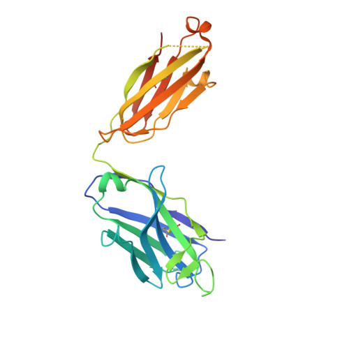
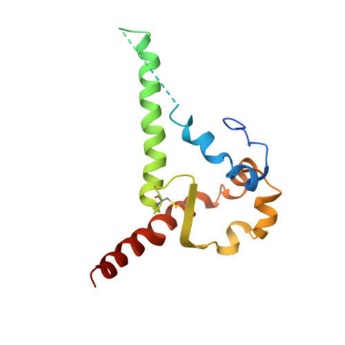
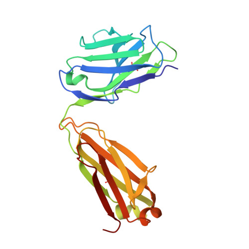
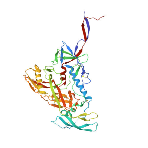
Broadly Neutralizing Antibody 8ANC195 Recognizes Closed and Open States of HIV-1 Env.

Scharf, L.Wang, H.Gao, H.Chen, S.McDowall, A.W.Bjorkman, P.J.

(2015) Cell 162: 1379-1390

  • DOI: https://doi.org/10.1016/j.cell.2015.08.035
  • Primary Citation of Related Structures:  
    5A7X, 5A8H, 5CJX

  • PubMed Abstract: 

    The HIV-1 envelope (Env) spike contains limited epitopes for broadly neutralizing antibodies (bNAbs); thus, most neutralizing antibodies are strain specific. The 8ANC195 epitope, defined by crystal and electron microscopy (EM) structures of bNAb 8ANC195 complexed with monomeric gp120 and trimeric Env, respectively, spans the gp120 and gp41 Env subunits. To investigate 8ANC195's gp41 epitope at higher resolution, we solved a 3.58 Å crystal structure of 8ANC195 complexed with fully glycosylated Env trimer, revealing 8ANC195 insertion into a glycan shield gap to contact gp120 and gp41 glycans and protein residues. To determine whether 8ANC195 recognizes the CD4-bound open Env conformation that leads to co-receptor binding and fusion, one of several known conformations of virion-associated Env, we solved EM structures of an Env/CD4/CD4-induced antibody/8ANC195 complex. 8ANC195 binding partially closed the CD4-bound trimer, confirming structural plasticity of Env by revealing a previously unseen conformation. 8ANC195's ability to bind different Env conformations suggests advantages for potential therapeutic applications.


  • Organizational Affiliation

    Division of Biology and Biological Engineering, California Institute of Technology, Pasadena, CA 91125, USA.


Macromolecules
Find similar proteins by:  (by identity cutoff)  |  3D Structure
Entity ID: 1
MoleculeChains Sequence LengthOrganismDetailsImage
8ANC195 G52K5 heavy chain, IG gamma-1 chainA,
D,
G [auth H]
244Homo sapiensMutation(s): 0 
Entity Groups  
Sequence Clusters30% Identity50% Identity70% Identity90% Identity95% Identity100% Identity
Sequence Annotations
Expand
  • Reference Sequence
Find similar proteins by:  (by identity cutoff)  |  3D Structure
Entity ID: 2
MoleculeChains Sequence LengthOrganismDetailsImage
BG505 Env gp41B,
H [auth J],
K [auth X]
153Human immunodeficiency virus 1Mutation(s): 2 
Gene Names: env
UniProt
Find proteins for Q2N0S6 (Human immunodeficiency virus 1)
Explore Q2N0S6 
Go to UniProtKB:  Q2N0S6
Entity Groups  
Sequence Clusters30% Identity50% Identity70% Identity90% Identity95% Identity100% Identity
UniProt GroupQ2N0S6
Sequence Annotations
Expand
  • Reference Sequence
Find similar proteins by:  (by identity cutoff)  |  3D Structure
Entity ID: 3
MoleculeChains Sequence LengthOrganismDetailsImage
8ANC195 G52K5 light chainC,
E,
J [auth L]
215Homo sapiensMutation(s): 0 
Entity Groups  
Sequence Clusters30% Identity50% Identity70% Identity90% Identity95% Identity100% Identity
Sequence Annotations
Expand
  • Reference Sequence
Find similar proteins by:  (by identity cutoff)  |  3D Structure
Entity ID: 4
MoleculeChains Sequence LengthOrganismDetailsImage
BG505 Env gp120F [auth G],
I [auth K],
L [auth Y]
479Human immunodeficiency virus 1Mutation(s): 2 
Gene Names: env
UniProt
Find proteins for Q2N0S6 (Human immunodeficiency virus 1)
Explore Q2N0S6 
Go to UniProtKB:  Q2N0S6
Entity Groups  
Sequence Clusters30% Identity50% Identity70% Identity90% Identity95% Identity100% Identity
UniProt GroupQ2N0S6
Sequence Annotations
Expand
  • Reference Sequence
Oligosaccharides

Help

Entity ID: 5
MoleculeChains Length2D Diagram Glycosylation3D Interactions
alpha-D-mannopyranose-(1-3)-[alpha-D-mannopyranose-(1-6)]beta-D-mannopyranose-(1-4)-2-acetamido-2-deoxy-beta-D-glucopyranose-(1-4)-2-acetamido-2-deoxy-beta-D-glucopyranoseBA [auth b],
M [auth F]
5N-Glycosylation
Glycosylation Resources
GlyTouCan:  G22768VO
GlyCosmos:  G22768VO
GlyGen:  G22768VO
Entity ID: 6
MoleculeChains Length2D Diagram Glycosylation3D Interactions
2-acetamido-2-deoxy-beta-D-glucopyranose-(1-4)-2-acetamido-2-deoxy-beta-D-glucopyranoseN [auth I],
O [auth M],
R [auth P],
S [auth Q],
X [auth V]
2N-Glycosylation
Glycosylation Resources
GlyTouCan:  G42666HT
GlyCosmos:  G42666HT
GlyGen:  G42666HT
Entity ID: 7
MoleculeChains Length2D Diagram Glycosylation3D Interactions
alpha-D-mannopyranose-(1-2)-alpha-D-mannopyranose-(1-2)-alpha-D-mannopyranose-(1-3)-[alpha-D-mannopyranose-(1-2)-alpha-D-mannopyranose-(1-6)-[alpha-D-mannopyranose-(1-3)]alpha-D-mannopyranose-(1-6)]beta-D-mannopyranose-(1-4)-2-acetamido-2-deoxy-beta-D-glucopyranose-(1-4)-2-acetamido-2-deoxy-beta-D-glucopyranoseP [auth N],
U [auth S],
Z
10N-Glycosylation
Glycosylation Resources
GlyTouCan:  G40702WU
GlyCosmos:  G40702WU
GlyGen:  G40702WU
Entity ID: 8
MoleculeChains Length2D Diagram Glycosylation3D Interactions
alpha-D-mannopyranose-(1-2)-alpha-D-mannopyranose-(1-3)-[alpha-D-mannopyranose-(1-6)]beta-D-mannopyranose-(1-4)-2-acetamido-2-deoxy-beta-D-glucopyranose-(1-4)-2-acetamido-2-deoxy-beta-D-glucopyranoseQ [auth O],
W [auth U]
6N-Glycosylation
Glycosylation Resources
GlyTouCan:  G56014GC
GlyCosmos:  G56014GC
GlyGen:  G56014GC
Entity ID: 9
MoleculeChains Length2D Diagram Glycosylation3D Interactions
alpha-D-mannopyranose-(1-3)-beta-D-mannopyranose-(1-4)-2-acetamido-2-deoxy-beta-D-glucopyranose-(1-4)-2-acetamido-2-deoxy-beta-D-glucopyranoseT [auth R],
Y [auth W]
4N-Glycosylation
Glycosylation Resources
GlyTouCan:  G81315DD
GlyCosmos:  G81315DD
GlyGen:  G81315DD
Entity ID: 10
MoleculeChains Length2D Diagram Glycosylation3D Interactions
beta-D-mannopyranose-(1-4)-2-acetamido-2-deoxy-beta-D-glucopyranose-(1-4)-2-acetamido-2-deoxy-beta-D-glucopyranoseAA [auth a],
V [auth T]
3N-Glycosylation
Glycosylation Resources
GlyTouCan:  G15407YE
GlyCosmos:  G15407YE
GlyGen:  G15407YE
Experimental Data & Validation

Experimental Data

  • Method: X-RAY DIFFRACTION
  • Resolution: 3.58 Å
  • R-Value Free: 0.286 
  • R-Value Work: 0.239 
  • R-Value Observed: 0.240 
  • Space Group: P 1 21 1
Unit Cell:
Length ( Å )Angle ( ˚ )
a = 117.739α = 90
b = 195.224β = 101.6
c = 119.094γ = 90
Software Package:
Software NamePurpose
PHENIXrefinement
Aimlessdata scaling
PHASERphasing
PDB_EXTRACTdata extraction
XDSdata reduction

Structure Validation

View Full Validation Report


Ligand Structure Quality Assessment 


Entry History & Funding Information

Deposition Data


Funding OrganizationLocationGrant Number
National Institutes of Health/National Institute Of Allergy and Infectious Diseases (NIH/NIAID)United StatesHIVRAD P01 AI100148
Bill & Melinda Gates FoundationUnited States1040753
American Cancer SocietyUnited StatesPF-13-076-01-MPC

Revision History  (Full details and data files)

  • Version 1.0: 2015-09-23
    Type: Initial release
  • Version 1.1: 2017-09-06
    Changes: Author supporting evidence, Derived calculations
  • Version 1.2: 2019-12-11
    Changes: Author supporting evidence
  • Version 2.0: 2020-07-29
    Type: Remediation
    Reason: Carbohydrate remediation
    Changes: Advisory, Atomic model, Data collection, Derived calculations, Structure summary
  • Version 2.1: 2023-09-27
    Changes: Data collection, Database references, Derived calculations, Refinement description, Structure summary