5ERK

X-ray structure of horse spleen apoferritin (control)


Experimental Data Snapshot

  • Method: X-RAY DIFFRACTION
  • Resolution: 2.00 Å
  • R-Value Free: 0.181 
  • R-Value Work: 0.143 
  • R-Value Observed: 0.145 

wwPDB Validation   3D Report Full Report


This is version 1.1 of the entry. See complete history


Literature

Cisplatin encapsulation within a ferritin nanocage: a high-resolution crystallographic study.

Pontillo, N.Pane, F.Messori, L.Amoresano, A.Merlino, A.

(2016) Chem Commun (Camb) 52: 4136-4139

  • DOI: https://doi.org/10.1039/c5cc10365g
  • Primary Citation of Related Structures:  
    5ERJ, 5ERK

  • PubMed Abstract: 

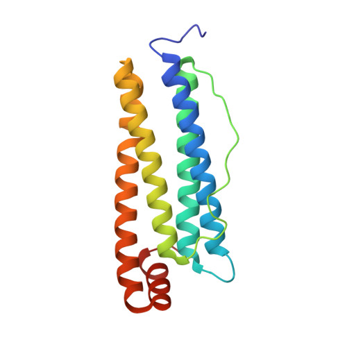
    Cisplatin (CDDP) can be encapsulated within the central cavity of reconstituted (apo)ferritin, (A)Ft, to form a drug-loaded protein of potential great interest for targeted cancer treatments. In this study, the interactions occurring between cisplatin and native horse spleen Ft in CDDP-encapsulated AFt are investigated by high-resolution X-ray crystallography. A protein bound Pt center is unambiguously identified in AFt subunits by comparative analysis of difference Fourier electron density maps and of anomalous dispersion data. Indeed, a [Pt(NH3)2H2O](2+) fragment is found coordinated to the His132 residue located on the inner surface of the large AFt spherical cage. Remarkably, Pt binding does not alter the overall physicochemical features (shape, volume, polarity/hydrophobicity and electrostatic potential) of the outer surface of the AFt nanocage. CDDP-encapsulated AFt appears to be an ideal nanocarrier for CDDP delivery to target sites, as it possesses high biocompatibility and can be internalized by receptor mediated endocytosis, thus carrying the drug to tumor tissue with higher selectivity than free CDDP.


  • Organizational Affiliation

    Department of Chemical Sciences, University of Naples Federico II, Complesso Universitario di Monte Sant'Angelo, Via Cintia, I-80126, Napoli, Italy. antonello.merlino@unina.it.


Macromolecules
Find similar proteins by:  (by identity cutoff)  |  3D Structure
Entity ID: 1
MoleculeChains Sequence LengthOrganismDetailsImage
Ferritin light chain174Equus caballusMutation(s): 0 
UniProt
Find proteins for P02791 (Equus caballus)
Explore P02791 
Go to UniProtKB:  P02791
Entity Groups  
Sequence Clusters30% Identity50% Identity70% Identity90% Identity95% Identity100% Identity
UniProt GroupP02791
Sequence Annotations
Expand
  • Reference Sequence
Small Molecules
Ligands 4 Unique
IDChains Name / Formula / InChI Key2D Diagram3D Interactions
CD
Query on CD

Download Ideal Coordinates CCD File 
B [auth A]
C [auth A]
D [auth A]
E [auth A]
F [auth A]
B [auth A],
C [auth A],
D [auth A],
E [auth A],
F [auth A],
G [auth A],
H [auth A],
I [auth A],
J [auth A],
K [auth A],
L [auth A],
M [auth A],
N [auth A]
CADMIUM ION
Cd
WLZRMCYVCSSEQC-UHFFFAOYSA-N
SO4
Query on SO4

Download Ideal Coordinates CCD File 
R [auth A]SULFATE ION
O4 S
QAOWNCQODCNURD-UHFFFAOYSA-L
GOL
Query on GOL

Download Ideal Coordinates CCD File 
S [auth A],
T [auth A]
GLYCEROL
C3 H8 O3
PEDCQBHIVMGVHV-UHFFFAOYSA-N
CL
Query on CL

Download Ideal Coordinates CCD File 
O [auth A],
P [auth A],
Q [auth A]
CHLORIDE ION
Cl
VEXZGXHMUGYJMC-UHFFFAOYSA-M
Experimental Data & Validation

Experimental Data

  • Method: X-RAY DIFFRACTION
  • Resolution: 2.00 Å
  • R-Value Free: 0.181 
  • R-Value Work: 0.143 
  • R-Value Observed: 0.145 
  • Space Group: F 4 3 2
Unit Cell:
Length ( Å )Angle ( ˚ )
a = 180.619α = 90
b = 180.619β = 90
c = 180.619γ = 90
Software Package:
Software NamePurpose
REFMACrefinement
XDSdata reduction
SCALAdata scaling
PHASERphasing

Structure Validation

View Full Validation Report


Entry History 

Deposition Data

Revision History  (Full details and data files)

  • Version 1.0: 2016-03-02
    Type: Initial release
  • Version 1.1: 2016-03-16
    Changes: Database references