5ET4

Structure of RNase A-K7H/R10H in complex with 3'-CMP


Experimental Data Snapshot

  • Method: X-RAY DIFFRACTION
  • Resolution: 2.10 Å
  • R-Value Free: 0.321 
  • R-Value Work: 0.224 
  • R-Value Observed: 0.229 

wwPDB Validation   3D Report Full Report


Ligand Structure Quality Assessment 


This is version 1.4 of the entry. See complete history


Literature

Characterization of an RNase with two catalytic centers. Human RNase6 catalytic and phosphate-binding site arrangement favors the endonuclease cleavage of polymeric substrates.

Prats-Ejarque, G.Blanco, J.A.Salazar, V.A.Nogues, V.M.Moussaoui, M.Boix, E.

(2019) Biochim Biophys Acta Gen Subj 1863: 105-117

  • DOI: https://doi.org/10.1016/j.bbagen.2018.09.021
  • Primary Citation of Related Structures:  
    5ET4, 5OAB, 5OGH, 6ENP

  • PubMed Abstract: 

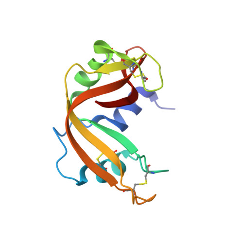
    Human RNase6 is a small cationic antimicrobial protein that belongs to the vertebrate RNaseA superfamily. All members share a common catalytic mechanism, which involves a conserved catalytic triad, constituted by two histidines and a lysine (His15/His122/Lys38 in RNase6 corresponding to His12/His119/Lys41 in RNaseA). Recently, our first crystal structure of human RNase6 identified an additional His pair (His36/His39) and suggested the presence of a secondary active site.


  • Organizational Affiliation

    Department of Biochemistry and Molecular Biology, Faculty of Biosciences, Universitat Autònoma de Barcelona, E-08193 Cerdanyola del Vallès, Spain. Electronic address: Guillem.Prats.Ejarque@uab.cat.


Macromolecules
Find similar proteins by:  (by identity cutoff)  |  3D Structure
Entity ID: 1
MoleculeChains Sequence LengthOrganismDetailsImage
Ribonuclease pancreatic
A, B, C, D
124Bos taurusMutation(s): 2 
Gene Names: RNASE1RNS1
EC: 3.1.27.5
UniProt
Find proteins for P61823 (Bos taurus)
Explore P61823 
Go to UniProtKB:  P61823
Entity Groups  
Sequence Clusters30% Identity50% Identity70% Identity90% Identity95% Identity100% Identity
UniProt GroupP61823
Sequence Annotations
Expand
  • Reference Sequence
Experimental Data & Validation

Experimental Data

  • Method: X-RAY DIFFRACTION
  • Resolution: 2.10 Å
  • R-Value Free: 0.321 
  • R-Value Work: 0.224 
  • R-Value Observed: 0.229 
  • Space Group: C 1 2 1
Unit Cell:
Length ( Å )Angle ( ˚ )
a = 160.366α = 90
b = 32.281β = 125.71
c = 106.963γ = 90
Software Package:
Software NamePurpose
PHENIXrefinement
SCALAdata scaling
PHASERphasing
XDSdata reduction

Structure Validation

View Full Validation Report


Ligand Structure Quality Assessment 


Entry History & Funding Information

Deposition Data


Funding OrganizationLocationGrant Number
Spain--

Revision History  (Full details and data files)

  • Version 1.0: 2016-11-30
    Type: Initial release
  • Version 1.1: 2018-01-24
    Changes: Source and taxonomy
  • Version 1.2: 2019-07-17
    Changes: Data collection
  • Version 1.3: 2021-02-24
    Changes: Database references
  • Version 1.4: 2024-01-10
    Changes: Data collection, Database references, Refinement description