5F3H

Structure of myostatin in complex with humanized RK35 antibody


Experimental Data Snapshot

  • Method: X-RAY DIFFRACTION
  • Resolution: 2.70 Å
  • R-Value Free: 0.270 
  • R-Value Work: 0.218 
  • R-Value Observed: 0.221 

wwPDB Validation   3D Report Full Report


This is version 1.2 of the entry. See complete history


Literature

Beyond CDR-grafting: Structure-guided humanization of framework and CDR regions of an anti-myostatin antibody.

Apgar, J.R.Mader, M.Agostinelli, R.Benard, S.Bialek, P.Johnson, M.Gao, Y.Krebs, M.Owens, J.Parris, K.St Andre, M.Svenson, K.Morris, C.Tchistiakova, L.

(2016) MAbs 8: 1302-1318

  • DOI: https://doi.org/10.1080/19420862.2016.1215786
  • Primary Citation of Related Structures:  
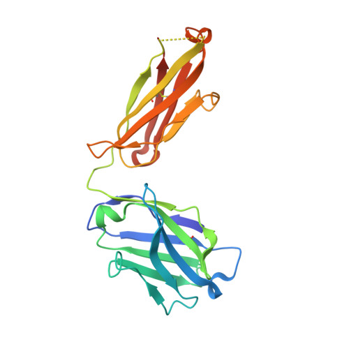
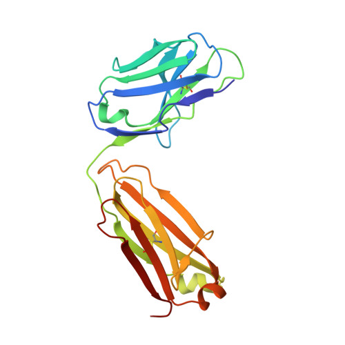
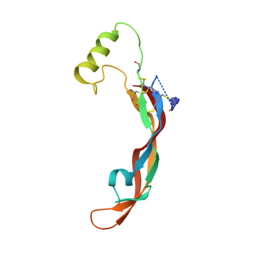
    5F3B, 5F3H

  • PubMed Abstract: 

    Antibodies are an important class of biotherapeutics that offer specificity to their antigen, long half-life, effector function interaction and good manufacturability. The immunogenicity of non-human-derived antibodies, which can be a major limitation to development, has been partially overcome by humanization through complementarity-determining region (CDR) grafting onto human acceptor frameworks. The retention of foreign content in the CDR regions, however, is still a potential immunogenic liability. Here, we describe the humanization of an anti-myostatin antibody utilizing a 2-step process of traditional CDR-grafting onto a human acceptor framework, followed by a structure-guided approach to further reduce the murine content of CDR-grafted antibodies. To accomplish this, we solved the co-crystal structures of myostatin with the chimeric (Protein Databank (PDB) id 5F3B) and CDR-grafted anti-myostatin antibody (PDB id 5F3H), allowing us to computationally predict the structurally important CDR residues as well as those making significant contacts with the antigen. Structure-based rational design enabled further germlining of the CDR-grafted antibody, reducing the murine content of the antibody without affecting antigen binding. The overall "humanness" was increased for both the light and heavy chain variable regions.


  • Organizational Affiliation

    a Biomedicine Design, Pfizer Inc. , Cambridge , MA , USA.


Macromolecules
Find similar proteins by:  (by identity cutoff)  |  3D Structure
Entity ID: 1
MoleculeChains Sequence LengthOrganismDetailsImage
humanized RK35 antibody heavy chain
A, C, E, G
221Mus musculusMutation(s): 0 
Entity Groups  
Sequence Clusters30% Identity50% Identity70% Identity90% Identity95% Identity100% Identity
Sequence Annotations
Expand
  • Reference Sequence
Find similar proteins by:  (by identity cutoff)  |  3D Structure
Entity ID: 2
MoleculeChains Sequence LengthOrganismDetailsImage
humanized RK35 antibody light chain
B, D, F, H
214Mus musculusMutation(s): 0 
Entity Groups  
Sequence Clusters30% Identity50% Identity70% Identity90% Identity95% Identity100% Identity
Sequence Annotations
Expand
  • Reference Sequence
Find similar proteins by:  (by identity cutoff)  |  3D Structure
Entity ID: 3
MoleculeChains Sequence LengthOrganismDetailsImage
Growth/differentiation factor 8
I, J, K, L
108Homo sapiensMutation(s): 0 
Gene Names: MSTNGDF8
UniProt & NIH Common Fund Data Resources
Find proteins for O14793 (Homo sapiens)
Explore O14793 
Go to UniProtKB:  O14793
PHAROS:  O14793
GTEx:  ENSG00000138379 
Entity Groups  
Sequence Clusters30% Identity50% Identity70% Identity90% Identity95% Identity100% Identity
UniProt GroupO14793
Sequence Annotations
Expand
  • Reference Sequence
Experimental Data & Validation

Experimental Data

  • Method: X-RAY DIFFRACTION
  • Resolution: 2.70 Å
  • R-Value Free: 0.270 
  • R-Value Work: 0.218 
  • R-Value Observed: 0.221 
  • Space Group: P 1
Unit Cell:
Length ( Å )Angle ( ˚ )
a = 75.138α = 88.33
b = 80.916β = 103.78
c = 101.401γ = 92.4
Software Package:
Software NamePurpose
PHENIXrefinement
SCALEPACKdata scaling
PDB_EXTRACTdata extraction
HKL-2000data reduction
AMoREphasing

Structure Validation

View Full Validation Report


Entry History 

Deposition Data

Revision History  (Full details and data files)

  • Version 1.0: 2016-09-28
    Type: Initial release
  • Version 1.1: 2016-10-12
    Changes: Database references
  • Version 1.2: 2023-09-27
    Changes: Data collection, Database references, Derived calculations, Refinement description