5HLU

X-ray crystal structure of met F43H/H64A sperm whale myoglobin in complex with nitric oxide


Experimental Data Snapshot

  • Method: X-RAY DIFFRACTION
  • Resolution: 1.50 Å
  • R-Value Free: 0.159 
  • R-Value Work: 0.141 
  • R-Value Observed: 0.142 

wwPDB Validation   3D Report Full Report


Ligand Structure Quality Assessment 


This is version 1.2 of the entry. See complete history


Literature

Regulating the nitrite reductase activity of myoglobin by redesigning the heme active center

Wu, L.B.Yuan, H.Gao, S.Q.You, Y.Nie, C.M.Wen, G.B.Lin, Y.W.Tan, X.

(2016) Nitric Oxide 57: 21-29

  • DOI: https://doi.org/10.1016/j.niox.2016.04.007
  • Primary Citation of Related Structures:  
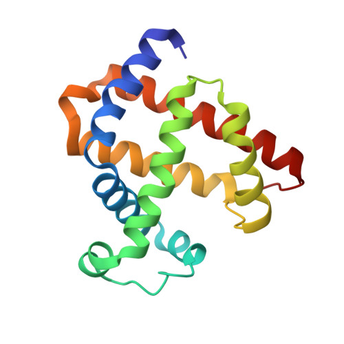
    5HLQ, 5HLU, 5HLX

  • PubMed Abstract: 

    Heme proteins perform diverse functions in living systems, of which nitrite reductase (NIR) activity receives much attention recently. In this study, to better understand the structural elements responsible for the NIR activity, we used myoglobin (Mb) as a model heme protein and redesigned the heme active center, by introducing one or two distal histidines, and by creating a channel to the heme center with removal of the native distal His64 gate (His to Ala mutation). UV-Vis kinetic studies, combined with EPR studies, showed that a single distal histidine with a suitable position to the heme iron, i.e., His43, is crucial for nitrite (NO2(-)) to nitric oxide (NO) reduction. Moreover, creation of a water channel to the heme center significantly enhanced the NIR activity compared to the corresponding mutant without the channel. In addition, X-ray crystallographic studies of F43H/H64A Mb and its complexes with NO2(-) or NO revealed a unique hydrogen-bonding network in the heme active center, as well as unique substrate and product binding models, providing valuable structural information for the enhanced NIR activity. These findings enriched our understanding of the structure and NIR activity relationship of heme proteins. The approach of creating a channel in this study is also useful for rational design of other functional heme proteins.


  • Organizational Affiliation

    School of Chemistry and Chemical Engineering, University of South China, Hengyang 421001, China.


Macromolecules
Find similar proteins by:  (by identity cutoff)  |  3D Structure
Entity ID: 1
MoleculeChains Sequence LengthOrganismDetailsImage
Myoglobin153Physeter catodonMutation(s): 2 
Gene Names: MB
UniProt
Find proteins for P02185 (Physeter macrocephalus)
Explore P02185 
Go to UniProtKB:  P02185
Entity Groups  
Sequence Clusters30% Identity50% Identity70% Identity90% Identity95% Identity100% Identity
UniProt GroupP02185
Sequence Annotations
Expand
  • Reference Sequence
Small Molecules
Ligands 2 Unique
IDChains Name / Formula / InChI Key2D Diagram3D Interactions
HEM
Query on HEM

Download Ideal Coordinates CCD File 
B [auth A]PROTOPORPHYRIN IX CONTAINING FE
C34 H32 Fe N4 O4
KABFMIBPWCXCRK-RGGAHWMASA-L
NO
Query on NO

Download Ideal Coordinates CCD File 
C [auth A]NITRIC OXIDE
N O
ODUCDPQEXGNKDN-UHFFFAOYSA-N
Experimental Data & Validation

Experimental Data

  • Method: X-RAY DIFFRACTION
  • Resolution: 1.50 Å
  • R-Value Free: 0.159 
  • R-Value Work: 0.141 
  • R-Value Observed: 0.142 
  • Space Group: P 21 21 21
Unit Cell:
Length ( Å )Angle ( ˚ )
a = 39.69α = 90
b = 49.528β = 90
c = 77.598γ = 90
Software Package:
Software NamePurpose
REFMACrefinement
HKL-3000data scaling
MOLREPphasing
HKL-3000data reduction

Structure Validation

View Full Validation Report


Ligand Structure Quality Assessment 


Entry History 

Deposition Data

  • Released Date: 2016-05-25 
  • Deposition Author(s): Yuan, H.

Revision History  (Full details and data files)

  • Version 1.0: 2016-05-25
    Type: Initial release
  • Version 1.1: 2016-12-21
    Changes: Structure summary
  • Version 1.2: 2023-11-08
    Changes: Data collection, Database references, Refinement description