5ITZ

Crystal structure of the SAC domain of CPAP in a complex with Tubulin and Darpin


Experimental Data Snapshot

  • Method: X-RAY DIFFRACTION
  • Resolution: 2.20 Å
  • R-Value Free: 0.217 
  • R-Value Work: 0.177 
  • R-Value Observed: 0.179 

wwPDB Validation   3D Report Full Report


Ligand Structure Quality Assessment 


This is version 1.3 of the entry. See complete history


Literature

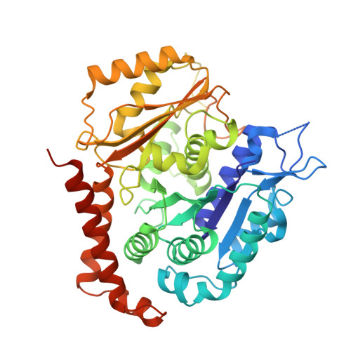
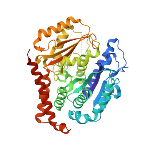
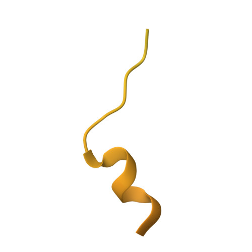
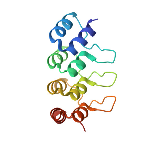
Centriolar CPAP/SAS-4 Imparts Slow Processive Microtubule Growth.

Sharma, A.Aher, A.Dynes, N.J.Frey, D.Katrukha, E.A.Jaussi, R.Grigoriev, I.Croisier, M.Kammerer, R.A.Akhmanova, A.Gonczy, P.Steinmetz, M.O.

(2016) Dev Cell 37: 362-376

  • DOI: https://doi.org/10.1016/j.devcel.2016.04.024
  • Primary Citation of Related Structures:  
    5ITZ

  • PubMed Abstract: 

    Centrioles are fundamental and evolutionarily conserved microtubule-based organelles whose assembly is characterized by microtubule growth rates that are orders of magnitude slower than those of cytoplasmic microtubules. Several centriolar proteins can interact with tubulin or microtubules, but how they ensure the exceptionally slow growth of centriolar microtubules has remained mysterious. Here, we bring together crystallographic, biophysical, and reconstitution assays to demonstrate that the human centriolar protein CPAP (SAS-4 in worms and flies) binds and "caps" microtubule plus ends by associating with a site of β-tubulin engaged in longitudinal tubulin-tubulin interactions. Strikingly, we uncover that CPAP activity dampens microtubule growth and stabilizes microtubules by inhibiting catastrophes and promoting rescues. We further establish that the capping function of CPAP is important to limit growth of centriolar microtubules in cells. Our results suggest that CPAP acts as a molecular lid that ensures slow assembly of centriolar microtubules and, thereby, contributes to organelle length control.


  • Organizational Affiliation

    Laboratory of Biomolecular Research, Department of Biology and Chemistry, Paul Scherrer Institut, 5232 Villigen PSI, Switzerland.


Macromolecules
Find similar proteins by:  (by identity cutoff)  |  3D Structure
Entity ID: 1
MoleculeChains Sequence LengthOrganismDetailsImage
Tubulin alpha-1B chain451Bos taurusMutation(s): 0 
UniProt
Find proteins for P81947 (Bos taurus)
Explore P81947 
Go to UniProtKB:  P81947
Entity Groups  
Sequence Clusters30% Identity50% Identity70% Identity90% Identity95% Identity100% Identity
UniProt GroupP81947
Sequence Annotations
Expand
  • Reference Sequence
Find similar proteins by:  (by identity cutoff)  |  3D Structure
Entity ID: 2
MoleculeChains Sequence LengthOrganismDetailsImage
Tubulin beta-2B chain445Bos taurusMutation(s): 0 
UniProt
Find proteins for Q6B856 (Bos taurus)
Explore Q6B856 
Go to UniProtKB:  Q6B856
Entity Groups  
Sequence Clusters30% Identity50% Identity70% Identity90% Identity95% Identity100% Identity
UniProt GroupQ6B856
Sequence Annotations
Expand
  • Reference Sequence
Find similar proteins by:  (by identity cutoff)  |  3D Structure
Entity ID: 3
MoleculeChains Sequence LengthOrganismDetailsImage
Centromere protein JC [auth D]129Homo sapiensMutation(s): 0 
Gene Names: CENPJCPAPLAPLIP1
UniProt & NIH Common Fund Data Resources
Find proteins for Q9HC77 (Homo sapiens)
Explore Q9HC77 
Go to UniProtKB:  Q9HC77
PHAROS:  Q9HC77
GTEx:  ENSG00000151849 
Entity Groups  
Sequence Clusters30% Identity50% Identity70% Identity90% Identity95% Identity100% Identity
UniProt GroupQ9HC77
Sequence Annotations
Expand
  • Reference Sequence
Find similar proteins by:  (by identity cutoff)  |  3D Structure
Entity ID: 4
MoleculeChains Sequence LengthOrganismDetailsImage
Designed ankyrin repeat protein (DARPIN) D1D [auth F]127synthetic constructMutation(s): 0 
Entity Groups  
Sequence Clusters30% Identity50% Identity70% Identity90% Identity95% Identity100% Identity
Sequence Annotations
Expand
  • Reference Sequence
Small Molecules
Ligands 4 Unique
IDChains Name / Formula / InChI Key2D Diagram3D Interactions
GTP
Query on GTP

Download Ideal Coordinates CCD File 
E [auth A]GUANOSINE-5'-TRIPHOSPHATE
C10 H16 N5 O14 P3
XKMLYUALXHKNFT-UUOKFMHZSA-N
GDP
Query on GDP

Download Ideal Coordinates CCD File 
G [auth B]GUANOSINE-5'-DIPHOSPHATE
C10 H15 N5 O11 P2
QGWNDRXFNXRZMB-UUOKFMHZSA-N
LOC
Query on LOC

Download Ideal Coordinates CCD File 
H [auth B]N-[(7S)-1,2,3,10-tetramethoxy-9-oxo-6,7-dihydro-5H-benzo[d]heptalen-7-yl]ethanamide
C22 H25 N O6
IAKHMKGGTNLKSZ-INIZCTEOSA-N
MG
Query on MG

Download Ideal Coordinates CCD File 
F [auth A]MAGNESIUM ION
Mg
JLVVSXFLKOJNIY-UHFFFAOYSA-N
Binding Affinity Annotations 
IDSourceBinding Affinity
LOC BindingDB:  5ITZ Ki: min: 1.6, max: 5750 (nM) from 4 assay(s)
Kd: min: 3700, max: 1.13e+4 (nM) from 2 assay(s)
IC50: min: 6.7, max: 2.93e+4 (nM) from 24 assay(s)
Experimental Data & Validation

Experimental Data

  • Method: X-RAY DIFFRACTION
  • Resolution: 2.20 Å
  • R-Value Free: 0.217 
  • R-Value Work: 0.177 
  • R-Value Observed: 0.179 
  • Space Group: P 1 21 1
Unit Cell:
Length ( Å )Angle ( ˚ )
a = 61.08α = 90
b = 85.34β = 91.77
c = 98.69γ = 90
Software Package:
Software NamePurpose
PHENIXrefinement
XDSdata reduction
XSCALEdata scaling
PHASERphasing

Structure Validation

View Full Validation Report


Ligand Structure Quality Assessment 


Entry History & Funding Information

Deposition Data


Funding OrganizationLocationGrant Number
Swiss National Science FoundationSwitzerland310030B_138659

Revision History  (Full details and data files)

  • Version 1.0: 2016-06-01
    Type: Initial release
  • Version 1.1: 2019-10-30
    Changes: Data collection, Source and taxonomy
  • Version 1.2: 2020-07-29
    Changes: Derived calculations, Structure summary
  • Version 1.3: 2024-01-10
    Changes: Data collection, Database references, Refinement description