5J7J

NMR Derived Structure of Ca2+ Calmodulin bound to Phosphorylated PSD-95


Experimental Data Snapshot

  • Method: SOLUTION NMR
  • Conformers Calculated: 200 
  • Conformers Submitted: 
  • Selection Criteria: structures with the lowest energy 

wwPDB Validation   3D Report Full Report


This is version 1.2 of the entry. See complete history


Literature

Ca2+/calmodulin binding to PSD-95 mediates homeostatic synaptic scaling down.

Chowdhury, D.Turner, M.Patriarchi, T.Hergarden, A.C.Anderson, D.Zhang, Y.Sun, J.Chen, C.Y.Ames, J.B.Hell, J.W.

(2018) EMBO J 37: 122-138

  • DOI: https://doi.org/10.15252/embj.201695829
  • Primary Citation of Related Structures:  
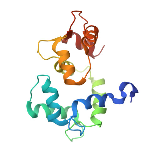
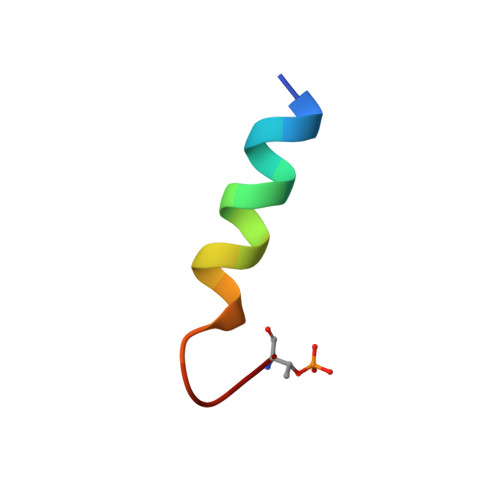
    5J7J

  • PubMed Abstract: 

    Postsynaptic density protein-95 (PSD-95) localizes AMPA-type glutamate receptors (AMPARs) to postsynaptic sites of glutamatergic synapses. Its postsynaptic displacement is necessary for loss of AMPARs during homeostatic scaling down of synapses. Here, we demonstrate that upon Ca 2+ influx, Ca 2+ /calmodulin (Ca 2+ /CaM) binding to the N-terminus of PSD-95 mediates postsynaptic loss of PSD-95 and AMPARs during homeostatic scaling down. Our NMR structural analysis identified E17 within the PSD-95 N-terminus as important for binding to Ca 2+ /CaM by interacting with R126 on CaM. Mutating E17 to R prevented homeostatic scaling down in primary hippocampal neurons, which is rescued via charge inversion by ectopic expression of CaM R 126E , as determined by analysis of miniature excitatory postsynaptic currents. Accordingly, increased binding of Ca 2+ /CaM to PSD-95 induced by a chronic increase in Ca 2+ influx is a critical molecular event in homeostatic downscaling of glutamatergic synaptic transmission.


  • Organizational Affiliation

    Department of Pharmacology, University of California, Davis, CA, USA.


Macromolecules
Find similar proteins by:  (by identity cutoff)  |  3D Structure
Entity ID: 1
MoleculeChains Sequence LengthOrganismDetailsImage
Calmodulin148Xenopus laevisMutation(s): 0 
Gene Names: calm1calm2
UniProt
Find proteins for P0DP33 (Xenopus laevis)
Explore P0DP33 
Go to UniProtKB:  P0DP33
Entity Groups  
Sequence Clusters30% Identity50% Identity70% Identity90% Identity95% Identity100% Identity
UniProt GroupP0DP33
Sequence Annotations
Expand
  • Reference Sequence

Find similar proteins by:  Sequence   |   3D Structure  

Entity ID: 2
MoleculeChains Sequence LengthOrganismDetailsImage
Disks large homolog 419Homo sapiensMutation(s): 0 
UniProt & NIH Common Fund Data Resources
Find proteins for P78352 (Homo sapiens)
Explore P78352 
Go to UniProtKB:  P78352
PHAROS:  P78352
GTEx:  ENSG00000132535 
Entity Groups  
Sequence Clusters30% Identity50% Identity70% Identity90% Identity95% Identity100% Identity
UniProt GroupP78352
Sequence Annotations
Expand
  • Reference Sequence
Small Molecules
Modified Residues  1 Unique
IDChains TypeFormula2D DiagramParent
TPO
Query on TPO
B
L-PEPTIDE LINKINGC4 H10 N O6 PTHR
Experimental Data & Validation

Experimental Data

  • Method: SOLUTION NMR
  • Conformers Calculated: 200 
  • Conformers Submitted: 
  • Selection Criteria: structures with the lowest energy 

Structure Validation

View Full Validation Report


Entry History & Funding Information

Deposition Data


Funding OrganizationLocationGrant Number
National Institutes of Health/National Institute of Mental Health (NIH/NIMH)United StatesMH097887
National Institutes of Health/National Eye Institute (NIH/NEI)United StatesEY012347

Revision History  (Full details and data files)

  • Version 1.0: 2017-10-25
    Type: Initial release
  • Version 1.1: 2019-05-08
    Changes: Data collection, Database references
  • Version 1.2: 2019-11-27
    Changes: Author supporting evidence, Data collection