5J8H

Structure of calmodulin in a complex with a peptide derived from a calmodulin-dependent kinase


Experimental Data Snapshot

  • Method: SOLUTION NMR
  • Conformers Calculated: 100 
  • Conformers Submitted: 20 
  • Selection Criteria: structures with the lowest energy 

wwPDB Validation   3D Report Full Report


This is version 1.4 of the entry. See complete history


Literature

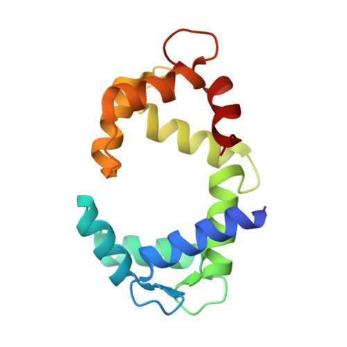
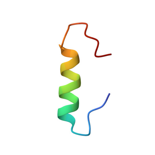
Structural Basis for the Recognition of Eukaryotic Elongation Factor 2 Kinase by Calmodulin.

Lee, K.Alphonse, S.Piserchio, A.Tavares, C.D.Giles, D.H.Wellmann, R.M.Dalby, K.N.Ghose, R.

(2016) Structure 24: 1441-1451

  • DOI: https://doi.org/10.1016/j.str.2016.06.015
  • Primary Citation of Related Structures:  
    5J8H

  • PubMed Abstract: 

    Binding of Ca(2+)-loaded calmodulin (CaM) activates eukaryotic elongation factor 2 kinase (eEF-2K) that phosphorylates eEF-2, its only known cellular target, leading to a decrease in global protein synthesis. Here, using an eEF-2K-derived peptide (eEF-2KCBD) that encodes the region necessary for its CaM-mediated activation, we provide a structural basis for their interaction. The striking feature of this association is the absence of Ca(2+) from the CaM C-lobe sites, even under high Ca(2+) conditions. eEF-2KCBD engages CaM largely through the C lobe of the latter in an anti-parallel 1-5-8 hydrophobic mode reinforced by a pair of unique electrostatic contacts. Sparse interactions of eEF-2KCBD with the CaM N lobe results in persisting inter-lobe mobility. A conserved eEF-2K residue (W85) anchors it to CaM by inserting into a deep hydrophobic cavity within the CaM C lobe. Mutation of this residue (W85S) substantially weakens interactions between full-length eEF-2K and CaM in vitro and reduces eEF-2 phosphorylation in cells.


  • Organizational Affiliation

    Department of Chemistry and Biochemistry, The City College of New York, New York, NY 10031, USA; Graduate Program in Biochemistry, The Graduate Center of CUNY, New York, NY 10016, USA.


Macromolecules
Find similar proteins by:  (by identity cutoff)  |  3D Structure
Entity ID: 1
MoleculeChains Sequence LengthOrganismDetailsImage
Calmodulin148Homo sapiensMutation(s): 0 
Gene Names: 
CALM1CALMCAMCAM1CALM2CAM2CAMBCALM3CALML2CAM3...
CALM1CALMCAMCAM1CALM2CAM2CAMBCALM3CALML2CAM3CAMCCAMIII

UniProt & NIH Common Fund Data Resources
Find proteins for P0DP23 (Homo sapiens)
Explore P0DP23 
Go to UniProtKB:  P0DP23
PHAROS:  P0DP23
GTEx:  ENSG00000198668 
Entity Groups  
Sequence Clusters30% Identity50% Identity70% Identity90% Identity95% Identity100% Identity
UniProt GroupP0DP23
Sequence Annotations
Expand
  • Reference Sequence
Find similar proteins by:  (by identity cutoff)  |  3D Structure
Entity ID: 2
MoleculeChains Sequence LengthOrganismDetailsImage
Eukaryotic elongation factor 2 kinase27Homo sapiensMutation(s): 0 
Gene Names: EEF2K
EC: 2.7.11.20
UniProt & NIH Common Fund Data Resources
Find proteins for O00418 (Homo sapiens)
Explore O00418 
Go to UniProtKB:  O00418
PHAROS:  O00418
GTEx:  ENSG00000103319 
Entity Groups  
Sequence Clusters30% Identity50% Identity70% Identity90% Identity95% Identity100% Identity
UniProt GroupO00418
Sequence Annotations
Expand
  • Reference Sequence
Small Molecules
Ligands 1 Unique
IDChains Name / Formula / InChI Key2D Diagram3D Interactions
CA
Query on CA

Download Ideal Coordinates CCD File 
C [auth A],
D [auth A]
CALCIUM ION
Ca
BHPQYMZQTOCNFJ-UHFFFAOYSA-N
Experimental Data & Validation

Experimental Data

  • Method: SOLUTION NMR
  • Conformers Calculated: 100 
  • Conformers Submitted: 20 
  • Selection Criteria: structures with the lowest energy 

Structure Validation

View Full Validation Report


Entry History & Funding Information

Deposition Data


Funding OrganizationLocationGrant Number
National Institutes of Health/National Institute of General Medical Sciences (NIH/NIGMS)United StatesGM084278
National Institutes of Health/National Institute on Minority Health and Health Disparities (NIH/NIMHD)United StatesG12 MD007603

Revision History  (Full details and data files)

  • Version 1.0: 2016-09-07
    Type: Initial release
  • Version 1.1: 2016-09-21
    Changes: Database references
  • Version 1.2: 2017-09-20
    Changes: Author supporting evidence, Database references, Structure summary
  • Version 1.3: 2019-12-18
    Changes: Author supporting evidence, Data collection
  • Version 1.4: 2023-06-14
    Changes: Database references, Other