5JCB

Microtubule depolymerizing agent podophyllotoxin derivative YJTSF1


Experimental Data Snapshot

  • Method: X-RAY DIFFRACTION
  • Resolution: 2.30 Å
  • R-Value Free: 0.200 
  • R-Value Work: 0.181 
  • R-Value Observed: 0.182 

wwPDB Validation   3D Report Full Report


Ligand Structure Quality Assessment 


This is version 1.2 of the entry. See complete history


Literature

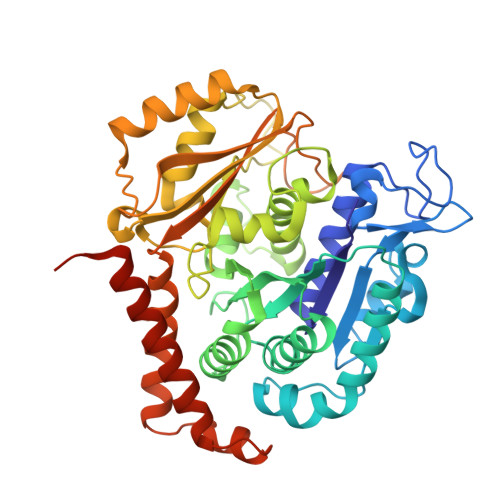
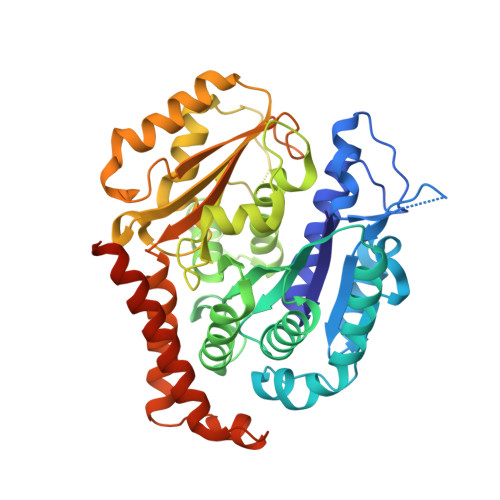
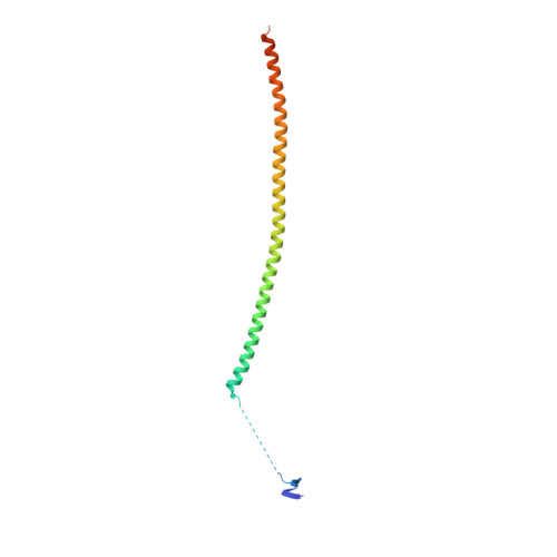
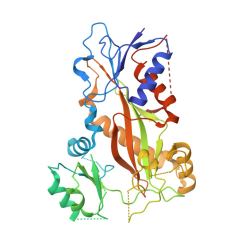
Structural Insights into the Inhibition of Tubulin by the Antitumor Agent 4 beta-(1,2,4-triazol-3-ylthio)-4-deoxypodophyllotoxin.

Zhao, W.Zhou, C.Guan, Z.Y.Yin, P.Chen, F.Tang, Y.J.

(2017) ACS Chem Biol 12: 746-752

  • DOI: https://doi.org/10.1021/acschembio.6b00842
  • Primary Citation of Related Structures:  
    5JCB

  • PubMed Abstract: 

    The colchicine domain is widely recognized as the binding site of microtubule depolymerization agents for anticancer drug design. Almost all of the drugs targeting the colchicine domain have been confirmed to bind to the tubulin β-subunit. Here we studied a crystal structure (2.3 Å) of the complex between tubulin and 4β-(1,2,4-triazol-3-ylthio)-4-deoxypodophyllotoxin (compound 1S) with superior antitumor activity, which was designed on the basis of the colchicine domain and synthesized in our previous work. A distinct binding model of the colchicine domain was found in the complexes of tubulin with compound 1S. From a comparison of the crystal structures of tubulin-compound 1S and tubulin-colchicine complexes, the side chains of the T7 loop of β-tubulin flip outward and the T5 loop of α-tubulin changes its conformation. It has been shown that the β-subunit T7 loop reversibly participates in resistance to straightening that opposes microtubule assembly by flipping in and out. Together with the biochemical results from compound 1S, the structural data highlight the main contributors in the α-subunits and the colchicine domain β-subunits: the dual-target binding sites in the α-T7 loop and β-H7-T7 loop of tubulin. Compound 1S can synchronously bind to αβ-tubulin. The structures also highlight common features for the design and development of novel potent microtubule destabilizing agents.


  • Organizational Affiliation

    National Key Laboratory of Agromicrobiology, College of Food Sciences and Technology, Huazhong Agricultural University , Wuhan 430070, China.


Macromolecules
Find similar proteins by:  (by identity cutoff)  |  3D Structure
Entity ID: 1
MoleculeChains Sequence LengthOrganismDetailsImage
Tubulin alpha-1B chain
A, C
451Sus scrofaMutation(s): 0 
UniProt
Find proteins for Q2XVP4 (Sus scrofa)
Explore Q2XVP4 
Go to UniProtKB:  Q2XVP4
Entity Groups  
Sequence Clusters30% Identity50% Identity70% Identity90% Identity95% Identity100% Identity
UniProt GroupQ2XVP4
Sequence Annotations
Expand
  • Reference Sequence
Find similar proteins by:  (by identity cutoff)  |  3D Structure
Entity ID: 2
MoleculeChains Sequence LengthOrganismDetailsImage
Tubulin beta chain
B, D
445Sus scrofaMutation(s): 0 
UniProt
Find proteins for P02554 (Sus scrofa)
Explore P02554 
Go to UniProtKB:  P02554
Entity Groups  
Sequence Clusters30% Identity50% Identity70% Identity90% Identity95% Identity100% Identity
UniProt GroupP02554
Sequence Annotations
Expand
  • Reference Sequence
Find similar proteins by:  (by identity cutoff)  |  3D Structure
Entity ID: 3
MoleculeChains Sequence LengthOrganismDetailsImage
Stathmin152Sus scrofaMutation(s): 0 
UniProt
Find proteins for F2Z508 (Sus scrofa)
Explore F2Z508 
Go to UniProtKB:  F2Z508
Entity Groups  
Sequence Clusters30% Identity50% Identity70% Identity90% Identity95% Identity100% Identity
UniProt GroupF2Z508
Sequence Annotations
Expand
  • Reference Sequence
Find similar proteins by:  (by identity cutoff)  |  3D Structure
Entity ID: 4
MoleculeChains Sequence LengthOrganismDetailsImage
Tubulin-Tyrosine Ligase388Gallus gallusMutation(s): 0 
Entity Groups  
Sequence Clusters30% Identity50% Identity70% Identity90% Identity95% Identity100% Identity
Sequence Annotations
Expand
  • Reference Sequence
Small Molecules
Ligands 11 Unique
IDChains Name / Formula / InChI Key2D Diagram3D Interactions
GTP
Query on GTP

Download Ideal Coordinates CCD File 
G [auth A],
T [auth C]
GUANOSINE-5'-TRIPHOSPHATE
C10 H16 N5 O14 P3
XKMLYUALXHKNFT-UUOKFMHZSA-N
ACP
Query on ACP

Download Ideal Coordinates CCD File 
JA [auth F]PHOSPHOMETHYLPHOSPHONIC ACID ADENYLATE ESTER
C11 H18 N5 O12 P3
UFZTZBNSLXELAL-IOSLPCCCSA-N
NV4
Query on NV4

Download Ideal Coordinates CCD File 
HA [auth D],
R [auth B]
(5R,5aR,8aS,9R)-9-[(4H-1,2,4-triazol-3-yl)sulfanyl]-5-(3,4,5-trimethoxyphenyl)-5,8,8a,9-tetrahydro-2H-furo[3',4':6,7]naphtho[2,3-d][1,3]dioxol-6(5aH)-one
C24 H23 N3 O7 S
WBKYKDGMCNMECE-QXHRYPSCSA-N
GDP
Query on GDP

Download Ideal Coordinates CCD File 
BA [auth D],
N [auth B]
GUANOSINE-5'-DIPHOSPHATE
C10 H15 N5 O11 P2
QGWNDRXFNXRZMB-UUOKFMHZSA-N
MES
Query on MES

Download Ideal Coordinates CCD File 
Q [auth B]2-(N-MORPHOLINO)-ETHANESULFONIC ACID
C6 H13 N O4 S
SXGZJKUKBWWHRA-UHFFFAOYSA-N
PEG
Query on PEG

Download Ideal Coordinates CCD File 
W [auth C]DI(HYDROXYETHYL)ETHER
C4 H10 O3
MTHSVFCYNBDYFN-UHFFFAOYSA-N
GOL
Query on GOL

Download Ideal Coordinates CCD File 
AA [auth C]
EA [auth D]
FA [auth D]
GA [auth D]
J [auth A]
AA [auth C],
EA [auth D],
FA [auth D],
GA [auth D],
J [auth A],
M [auth A],
S [auth C],
V [auth C]
GLYCEROL
C3 H8 O3
PEDCQBHIVMGVHV-UHFFFAOYSA-N
IMD
Query on IMD

Download Ideal Coordinates CCD File 
K [auth A],
Z [auth C]
IMIDAZOLE
C3 H5 N2
RAXXELZNTBOGNW-UHFFFAOYSA-O
CA
Query on CA

Download Ideal Coordinates CCD File 
L [auth A],
Y [auth C]
CALCIUM ION
Ca
BHPQYMZQTOCNFJ-UHFFFAOYSA-N
MG
Query on MG

Download Ideal Coordinates CCD File 
CA [auth D]
DA [auth D]
H [auth A]
I [auth A]
IA [auth F]
CA [auth D],
DA [auth D],
H [auth A],
I [auth A],
IA [auth F],
O [auth B],
U [auth C]
MAGNESIUM ION
Mg
JLVVSXFLKOJNIY-UHFFFAOYSA-N
NA
Query on NA

Download Ideal Coordinates CCD File 
P [auth B],
X [auth C]
SODIUM ION
Na
FKNQFGJONOIPTF-UHFFFAOYSA-N
Experimental Data & Validation

Experimental Data

  • Method: X-RAY DIFFRACTION
  • Resolution: 2.30 Å
  • R-Value Free: 0.200 
  • R-Value Work: 0.181 
  • R-Value Observed: 0.182 
  • Space Group: P 21 21 21
Unit Cell:
Length ( Å )Angle ( ˚ )
a = 104.357α = 90
b = 157.252β = 90
c = 179.631γ = 90
Software Package:
Software NamePurpose
PHENIXrefinement
HKL-2000data collection
HKL-2000data scaling
PHASERphasing

Structure Validation

View Full Validation Report


Ligand Structure Quality Assessment 


Entry History 

Deposition Data

Revision History  (Full details and data files)

  • Version 1.0: 2017-09-27
    Type: Initial release
  • Version 1.1: 2017-11-08
    Changes: Source and taxonomy
  • Version 1.2: 2023-11-08
    Changes: Data collection, Database references, Derived calculations, Refinement description