5JD2

SFX structure of corestreptavidin-selenobiotin complex


Experimental Data Snapshot

  • Method: X-RAY DIFFRACTION
  • Resolution: 1.90 Å
  • R-Value Free: 0.200 
  • R-Value Work: 0.165 
  • R-Value Observed: 0.166 

wwPDB Validation   3D Report Full Report


This is version 1.5 of the entry. See complete history


Literature

Selenium single-wavelength anomalous diffraction de novo phasing using an X-ray-free electron laser.

Hunter, M.S.Yoon, C.H.DeMirci, H.Sierra, R.G.Dao, E.H.Ahmadi, R.Aksit, F.Aquila, A.L.Ciftci, H.Guillet, S.Hayes, M.J.Lane, T.J.Liang, M.Lundstrom, U.Koglin, J.E.Mgbam, P.Rao, Y.Zhang, L.Wakatsuki, S.Holton, J.M.Boutet, S.

(2016) Nat Commun 7: 13388-13388

  • DOI: https://doi.org/10.1038/ncomms13388
  • Primary Citation of Related Structures:  
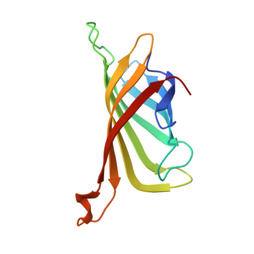
    5JD2

  • PubMed Abstract: 

    Structural information about biological macromolecules near the atomic scale provides important insight into the functions of these molecules. To date, X-ray crystallography has been the predominant method used for macromolecular structure determination. However, challenges exist when solving structures with X-rays, including the phase problem and radiation damage. X-ray-free electron lasers (X-ray FELs) have enabled collection of diffraction information before the onset of radiation damage, yet the majority of structures solved at X-ray FELs have been phased using external information via molecular replacement. De novo phasing at X-ray FELs has proven challenging due in part to per-pulse variations in intensity and wavelength. Here we report the solution of a selenobiotinyl-streptavidin structure using phases obtained by the anomalous diffraction of selenium measured at a single wavelength (Se-SAD) at the Linac Coherent Light Source. Our results demonstrate Se-SAD, routinely employed at synchrotrons for novel structure determination, is now possible at X-ray FELs.


  • Organizational Affiliation

    Linac Coherent Light Source, SLAC National Accelerator Laboratory, Menlo Park, California 94025, USA.


Macromolecules
Find similar proteins by:  (by identity cutoff)  |  3D Structure
Entity ID: 1
MoleculeChains Sequence LengthOrganismDetailsImage
Streptavidin
A, B, C, D
120Streptomyces avidiniiMutation(s): 0 
UniProt
Find proteins for P22629 (Streptomyces avidinii)
Explore P22629 
Go to UniProtKB:  P22629
Entity Groups  
Sequence Clusters30% Identity50% Identity70% Identity90% Identity95% Identity100% Identity
UniProt GroupP22629
Sequence Annotations
Expand
  • Reference Sequence
Experimental Data & Validation

Experimental Data

  • Method: X-RAY DIFFRACTION
  • Resolution: 1.90 Å
  • R-Value Free: 0.200 
  • R-Value Work: 0.165 
  • R-Value Observed: 0.166 
  • Space Group: P 1 21 1
Unit Cell:
Length ( Å )Angle ( ˚ )
a = 50.84α = 90
b = 98.52β = 112.38
c = 53.43γ = 90
Software Package:
Software NamePurpose
DMphasing
PHENIXrefinement
PDB_EXTRACTdata extraction
CrystFELdata reduction
CrystFELdata scaling

Structure Validation

View Full Validation Report


Entry History & Funding Information

Deposition Data


Funding OrganizationLocationGrant Number
Department of Energy (DOE, United States)United States--

Revision History  (Full details and data files)

  • Version 1.0: 2016-11-16
    Type: Initial release
  • Version 1.1: 2017-09-06
    Changes: Advisory, Author supporting evidence, Derived calculations
  • Version 1.2: 2017-11-22
    Changes: Refinement description
  • Version 1.3: 2018-02-14
    Changes: Data collection
  • Version 1.4: 2019-12-04
    Changes: Author supporting evidence
  • Version 1.5: 2024-03-06
    Changes: Advisory, Data collection, Database references