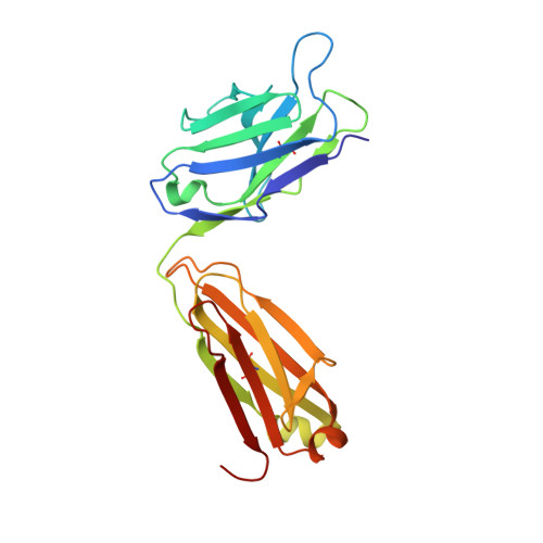
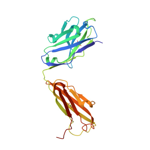
5JUE

Crystal Structure of UIC2 Fab


Experimental Data Snapshot

  • Method: X-RAY DIFFRACTION
  • Resolution: 1.65 Å
  • R-Value Free: 0.203 
  • R-Value Work: 0.165 
  • R-Value Observed: 0.166 

wwPDB Validation   3D Report Full Report


This is version 1.1 of the entry. See complete history


Literature

Crystal structure of the antigen-binding fragment of a monoclonal antibody specific for the multidrug-resistance-linked ABC transporter human P-glycoprotein.

Esser, L.Shukla, S.Zhou, F.Ambudkar, S.V.Xia, D.

(2016) Acta Crystallogr F Struct Biol Commun 72: 636-641

  • DOI: https://doi.org/10.1107/S2053230X16009778
  • Primary Citation of Related Structures:  
    5JUE

  • PubMed Abstract: 

    P-glycoprotein (P-gp) is a polyspecific ATP-dependent transporter linked to multidrug resistance in cancers that plays important roles in the pharmacokinetics of a large number of drugs. The drug-resistance phenotype of P-gp can be modulated by the monoclonal antibody UIC2, which specifically recognizes human P-gp in a conformation-dependent manner. Here, the purification, sequence determination and high-resolution structure of the Fab fragment of UIC2 (UIC2/Fab) are reported. Purified UIC2/Fab binds human P-gp with a 1:1 stoichiometry. Crystals of UIC2/Fab are triclinic (space group P1), with unit-cell parameters a = 40.67, b = 44.91, c = 58.09 Å, α = 97.62, β = 99.10, γ = 94.09°, and diffracted X-rays to 1.6 Å resolution. The structure was determined by molecular replacement and refined to 1.65 Å resolution. The asymmetric unit contains one molecule of UIC2/Fab, which exhibits a positively charged antigen-binding surface, suggesting that it might recognize an oppositely charged extracellular epitope of P-gp.


  • Organizational Affiliation

    National Cancer Institute, National Institutes of Health, 37 Convent Drive, Building 37, Bethesda, MD 20892, USA.


Macromolecules
Find similar proteins by:  (by identity cutoff)  |  3D Structure
Entity ID: 1
MoleculeChains Sequence LengthOrganismDetailsImage
light chain of UIC2 FabA [auth L]220Mus musculusMutation(s): 0 
Entity Groups  
Sequence Clusters30% Identity50% Identity70% Identity90% Identity95% Identity100% Identity
Sequence Annotations
Expand
  • Reference Sequence
Find similar proteins by:  (by identity cutoff)  |  3D Structure
Entity ID: 2
MoleculeChains Sequence LengthOrganismDetailsImage
heavy chain of UIC2 FabB [auth H]225Mus musculusMutation(s): 0 
Entity Groups  
Sequence Clusters30% Identity50% Identity70% Identity90% Identity95% Identity100% Identity
Sequence Annotations
Expand
  • Reference Sequence
Experimental Data & Validation

Experimental Data

  • Method: X-RAY DIFFRACTION
  • Resolution: 1.65 Å
  • R-Value Free: 0.203 
  • R-Value Work: 0.165 
  • R-Value Observed: 0.166 
  • Space Group: P 1
Unit Cell:
Length ( Å )Angle ( ˚ )
a = 40.673α = 97.62
b = 44.909β = 99.1
c = 58.094γ = 94.09
Software Package:
Software NamePurpose
PHENIXrefinement
DENZOdata reduction
SCALEPACKdata scaling
MOLREPphasing
PDB_EXTRACTdata extraction

Structure Validation

View Full Validation Report


Entry History 

Deposition Data

Revision History  (Full details and data files)

  • Version 1.0: 2016-08-31
    Type: Initial release
  • Version 1.1: 2023-09-27
    Changes: Data collection, Database references, Derived calculations, Refinement description