5K8Q

Crystal Structure of Calcium-loaded Calmodulin in complex with STRA6 CaMBP2-site peptide.


Experimental Data Snapshot

  • Method: X-RAY DIFFRACTION
  • Resolution: 1.74 Å
  • R-Value Free: 0.267 
  • R-Value Work: 0.227 
  • R-Value Observed: 0.229 

wwPDB Validation   3D Report Full Report


This is version 1.4 of the entry. See complete history


Literature

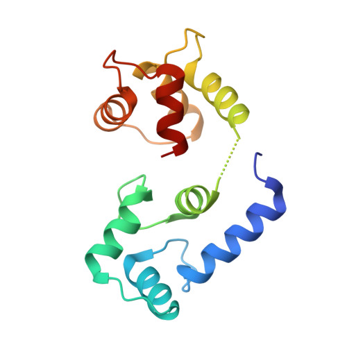
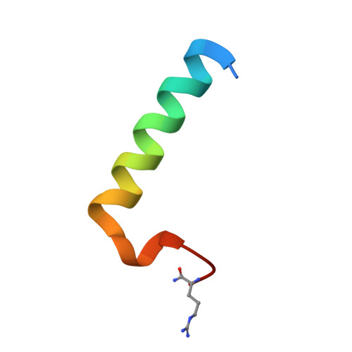
Structure of the STRA6 receptor for retinol uptake.

Chen, Y.Clarke, O.B.Kim, J.Stowe, S.Kim, Y.K.Assur, Z.Cavalier, M.Godoy-Ruiz, R.von Alpen, D.C.Manzini, C.Blaner, W.S.Frank, J.Quadro, L.Weber, D.J.Shapiro, L.Hendrickson, W.A.Mancia, F.

(2016) Science 353

  • DOI: https://doi.org/10.1126/science.aad8266
  • Primary Citation of Related Structures:  
    5K8Q, 5SY1

  • PubMed Abstract: 

    Vitamin A homeostasis is critical to normal cellular function. Retinol-binding protein (RBP) is the sole specific carrier in the bloodstream for hydrophobic retinol, the main form in which vitamin A is transported. The integral membrane receptor STRA6 mediates cellular uptake of vitamin A by recognizing RBP-retinol to trigger release and internalization of retinol. We present the structure of zebrafish STRA6 determined to 3.9-angstrom resolution by single-particle cryo-electron microscopy. STRA6 has one intramembrane and nine transmembrane helices in an intricate dimeric assembly. Unexpectedly, calmodulin is bound tightly to STRA6 in a noncanonical arrangement. Residues involved with RBP binding map to an archlike structure that covers a deep lipophilic cleft. This cleft is open to the membrane, suggesting a possible mode for internalization of retinol through direct diffusion into the lipid bilayer.


  • Organizational Affiliation

    Department of Physiology and Cellular Biophysics, Columbia University, New York, NY 10032, USA.


Macromolecules
Find similar proteins by:  (by identity cutoff)  |  3D Structure
Entity ID: 1
MoleculeChains Sequence LengthOrganismDetailsImage
Calmodulin149Homo sapiensMutation(s): 0 
Gene Names: 
CALM1CALMCAMCAM1CALM2CAM2CAMBCALM3CALML2CAM3...
CALM1CALMCAMCAM1CALM2CAM2CAMBCALM3CALML2CAM3CAMCCAMIII

UniProt & NIH Common Fund Data Resources
Find proteins for P0DP23 (Homo sapiens)
Explore P0DP23 
Go to UniProtKB:  P0DP23
PHAROS:  P0DP23
GTEx:  ENSG00000198668 
Entity Groups  
Sequence Clusters30% Identity50% Identity70% Identity90% Identity95% Identity100% Identity
UniProt GroupP0DP23
Sequence Annotations
Expand
  • Reference Sequence
Find similar proteins by:  (by identity cutoff)  |  3D Structure
Entity ID: 2
MoleculeChains Sequence LengthOrganismDetailsImage
Zgc:13668929Danio rerioMutation(s): 2 
UniProt
Find proteins for F1RAX4 (Danio rerio)
Explore F1RAX4 
Go to UniProtKB:  F1RAX4
Entity Groups  
Sequence Clusters30% Identity50% Identity70% Identity90% Identity95% Identity100% Identity
UniProt GroupF1RAX4
Sequence Annotations
Expand
  • Reference Sequence
Experimental Data & Validation

Experimental Data

  • Method: X-RAY DIFFRACTION
  • Resolution: 1.74 Å
  • R-Value Free: 0.267 
  • R-Value Work: 0.227 
  • R-Value Observed: 0.229 
  • Space Group: C 1 2 1
Unit Cell:
Length ( Å )Angle ( ˚ )
a = 156.51α = 90
b = 37.23β = 94.35
c = 35.73γ = 90
Software Package:
Software NamePurpose
PHENIXrefinement
iMOSFLMdata reduction
Aimlessdata scaling
PHASERphasing

Structure Validation

View Full Validation Report


Entry History & Funding Information

Deposition Data


Funding OrganizationLocationGrant Number
National Institutes of Health/National Institute of General Medical Sciences (NIH/NIGMS)United StatesU54 GM095315

Revision History  (Full details and data files)

  • Version 1.0: 2016-08-24
    Type: Initial release
  • Version 1.1: 2016-09-07
    Changes: Database references
  • Version 1.2: 2017-09-13
    Changes: Author supporting evidence, Database references, Derived calculations
  • Version 1.3: 2019-12-25
    Changes: Author supporting evidence
  • Version 1.4: 2024-04-03
    Changes: Data collection, Database references, Derived calculations, Refinement description