5LXT

Tubulin-Discodermolide complex


Experimental Data Snapshot

  • Method: X-RAY DIFFRACTION
  • Resolution: 1.90 Å
  • R-Value Free: 0.208 
  • R-Value Work: 0.173 
  • R-Value Observed: 0.175 

wwPDB Validation   3D Report Full Report


Ligand Structure Quality Assessment 


This is version 1.4 of the entry. See complete history


Literature

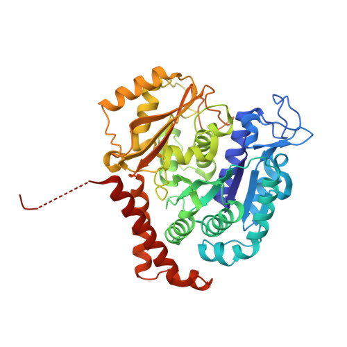
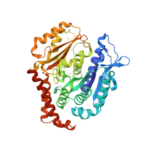
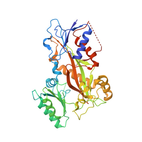
Structural Basis of Microtubule Stabilization by Discodermolide.

Prota, A.E.Bargsten, K.Redondo-Horcajo, M.Smith, A.B.Yang, C.H.McDaid, H.M.Paterson, I.Horwitz, S.B.Fernando Diaz, J.Steinmetz, M.O.

(2017) Chembiochem 18: 905-909

  • DOI: https://doi.org/10.1002/cbic.201600696
  • Primary Citation of Related Structures:  
    5LXS, 5LXT

  • PubMed Abstract: 

    Microtubule-stabilizing agents (MSAs) are widely used in chemotherapy. Using X-ray crystallography we elucidated the detailed binding modes of two potent MSAs, (+)-discodermolide (DDM) and the DDM-paclitaxel hybrid KS-1-199-32, in the taxane pocket of β-tubulin. The two compounds bind in a very similar hairpin conformation, as previously observed in solution. However, they stabilize the M-loop of β-tubulin differently: KS-1-199-32 induces an M-loop helical conformation that is not observed for DDM. In the context of the microtubule structure, both MSAs connect the β-tubulin helices H6 and H7 and loop S9-S10 with the M-loop. This is similar to the structural effects elicited by epothilone A, but distinct from paclitaxel. Together, our data reveal differential binding mechanisms of DDM and KS-1-199-32 on tubulin.


  • Organizational Affiliation

    Laboratory of Biomolecular Research, Department of Biology and Chemistry, Paul Scherrer Institut, OFLC/111, 5232, Villigen PSI, Switzerland.


Macromolecules
Find similar proteins by:  (by identity cutoff)  |  3D Structure
Entity ID: 1
MoleculeChains Sequence LengthOrganismDetailsImage
Tubulin alpha-1B chain
A, C
451Bos taurusMutation(s): 0 
UniProt
Find proteins for P81947 (Bos taurus)
Explore P81947 
Go to UniProtKB:  P81947
Entity Groups  
Sequence Clusters30% Identity50% Identity70% Identity90% Identity95% Identity100% Identity
UniProt GroupP81947
Sequence Annotations
Expand
  • Reference Sequence
Find similar proteins by:  (by identity cutoff)  |  3D Structure
Entity ID: 2
MoleculeChains Sequence LengthOrganismDetailsImage
Tubulin beta-2B chain
B, D
445Bos taurusMutation(s): 0 
UniProt
Find proteins for Q6B856 (Bos taurus)
Explore Q6B856 
Go to UniProtKB:  Q6B856
Entity Groups  
Sequence Clusters30% Identity50% Identity70% Identity90% Identity95% Identity100% Identity
UniProt GroupQ6B856
Sequence Annotations
Expand
  • Reference Sequence
Find similar proteins by:  (by identity cutoff)  |  3D Structure
Entity ID: 3
MoleculeChains Sequence LengthOrganismDetailsImage
Stathmin-4143Rattus norvegicusMutation(s): 0 
Gene Names: Stmn4
UniProt
Find proteins for P63043 (Rattus norvegicus)
Explore P63043 
Go to UniProtKB:  P63043
Entity Groups  
Sequence Clusters30% Identity50% Identity70% Identity90% Identity95% Identity100% Identity
UniProt GroupP63043
Sequence Annotations
Expand
  • Reference Sequence
Find similar proteins by:  (by identity cutoff)  |  3D Structure
Entity ID: 4
MoleculeChains Sequence LengthOrganismDetailsImage
TUBULIN-TYROSINE LIGASE384Gallus gallusMutation(s): 0 
Gene Names: TTL
Entity Groups  
Sequence Clusters30% Identity50% Identity70% Identity90% Identity95% Identity100% Identity
Sequence Annotations
Expand
  • Reference Sequence
Small Molecules
Ligands 7 Unique
IDChains Name / Formula / InChI Key2D Diagram3D Interactions
7AK
Query on 7AK

Download Ideal Coordinates CCD File 
O [auth B],
U [auth D]
(+)-Discodermolide
C33 H55 N O8
AADVCYNFEREWOS-OBRABYBLSA-N
GTP
Query on GTP

Download Ideal Coordinates CCD File 
G [auth A],
P [auth C]
GUANOSINE-5'-TRIPHOSPHATE
C10 H16 N5 O14 P3
XKMLYUALXHKNFT-UUOKFMHZSA-N
ACP
Query on ACP

Download Ideal Coordinates CCD File 
V [auth F]PHOSPHOMETHYLPHOSPHONIC ACID ADENYLATE ESTER
C11 H18 N5 O12 P3
UFZTZBNSLXELAL-IOSLPCCCSA-N
GDP
Query on GDP

Download Ideal Coordinates CCD File 
K [auth B],
S [auth D]
GUANOSINE-5'-DIPHOSPHATE
C10 H15 N5 O11 P2
QGWNDRXFNXRZMB-UUOKFMHZSA-N
MES
Query on MES

Download Ideal Coordinates CCD File 
N [auth B]2-(N-MORPHOLINO)-ETHANESULFONIC ACID
C6 H13 N O4 S
SXGZJKUKBWWHRA-UHFFFAOYSA-N
CA
Query on CA

Download Ideal Coordinates CCD File 
I [auth A],
J [auth A],
M [auth B],
R [auth C]
CALCIUM ION
Ca
BHPQYMZQTOCNFJ-UHFFFAOYSA-N
MG
Query on MG

Download Ideal Coordinates CCD File 
H [auth A],
L [auth B],
Q [auth C],
T [auth D],
W [auth F]
MAGNESIUM ION
Mg
JLVVSXFLKOJNIY-UHFFFAOYSA-N
Binding Affinity Annotations 
IDSourceBinding Affinity
7AK Binding MOAD:  5LXT Ka: 8.72e+9 (M^-1) from 1 assay(s)
Experimental Data & Validation

Experimental Data

  • Method: X-RAY DIFFRACTION
  • Resolution: 1.90 Å
  • R-Value Free: 0.208 
  • R-Value Work: 0.173 
  • R-Value Observed: 0.175 
  • Space Group: P 21 21 21
Unit Cell:
Length ( Å )Angle ( ˚ )
a = 104.5α = 90
b = 157.79β = 90
c = 180.23γ = 90
Software Package:
Software NamePurpose
PHENIXrefinement
XDSdata reduction
XSCALEdata scaling
PHENIXphasing

Structure Validation

View Full Validation Report


Ligand Structure Quality Assessment 


Entry History & Funding Information

Deposition Data


Funding OrganizationLocationGrant Number
Swiss National Science FoundationSwitzerland310030B_138659 and 31003A_166608
Spanish Ministry of Economy and CompetitivenessSpainBIO2013-42984-R and S2010/BMD-2457 BIPEDD2
National Institutes of Health/National Cancer Institute (NIH/NCI)United StatesCA077263
Breast Cancer Research FoundationUnited States--

Revision History  (Full details and data files)

  • Version 1.0: 2017-03-01
    Type: Initial release
  • Version 1.1: 2017-06-07
    Changes: Database references
  • Version 1.2: 2017-09-13
    Changes: Author supporting evidence
  • Version 1.3: 2022-03-30
    Changes: Author supporting evidence, Database references, Derived calculations
  • Version 1.4: 2024-01-17
    Changes: Data collection, Refinement description