5M78

Human Carbonic Anhydrase II in complex with fragment-like inhibitor.


Experimental Data Snapshot

  • Method: X-RAY DIFFRACTION
  • Resolution: 1.08 Å
  • R-Value Free: 0.144 
  • R-Value Work: 0.125 
  • R-Value Observed: 0.126 

wwPDB Validation   3D Report Full Report


Ligand Structure Quality Assessment 


This is version 1.2 of the entry. See complete history


Literature

A Proof-of-Concept Fragment Screening of a Hit-Validated 96-Compounds Library against Human Carbonic Anhydrase II.

Glockner, S.Heine, A.Klebe, G.

(2020) Biomolecules 10

  • DOI: https://doi.org/10.3390/biom10040518
  • Primary Citation of Related Structures:  
    5M78, 6RM1, 6RMP, 6S9Z, 6SAC, 6SAS, 6SAY, 6SB7, 6SDJ

  • PubMed Abstract: 

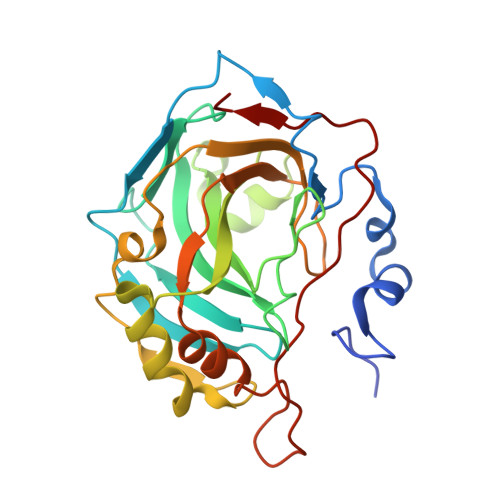
    Fragment screening is a powerful tool to identify and characterize binding pockets in proteins. We herein present the results of a proof-of-concept screening campaign of a versatile 96-entry fragment library from our laboratory against the drug target and model protein human carbonic anhydrase II. The screening revealed a novel chemotype for carbonic anhydrase inhibition, as well as less common non-covalent interaction types and unexpected covalent linkages. Lastly, different runs of the PanDDA tool reveal a practical hint for its application.


  • Organizational Affiliation

    Institute for Pharmaceutical Chemistry, Philipps-Universität Marburg, Marbacher Weg 6, 35037 Marburg, Germany.


Macromolecules
Find similar proteins by:  (by identity cutoff)  |  3D Structure
Entity ID: 1
MoleculeChains Sequence LengthOrganismDetailsImage
Carbonic anhydrase 2265Homo sapiensMutation(s): 0 
Gene Names: CA2
EC: 4.2.1.1
UniProt & NIH Common Fund Data Resources
Find proteins for P00918 (Homo sapiens)
Explore P00918 
Go to UniProtKB:  P00918
PHAROS:  P00918
GTEx:  ENSG00000104267 
Entity Groups  
Sequence Clusters30% Identity50% Identity70% Identity90% Identity95% Identity100% Identity
UniProt GroupP00918
Sequence Annotations
Expand
  • Reference Sequence
Small Molecules
Binding Affinity Annotations 
IDSourceBinding Affinity
SAL BindingDB:  5M78 Ki: min: 7100, max: 7.40e+5 (nM) from 3 assay(s)
IC50: 6.80e+5 (nM) from 1 assay(s)
Experimental Data & Validation

Experimental Data

  • Method: X-RAY DIFFRACTION
  • Resolution: 1.08 Å
  • R-Value Free: 0.144 
  • R-Value Work: 0.125 
  • R-Value Observed: 0.126 
  • Space Group: P 1 21 1
Unit Cell:
Length ( Å )Angle ( ˚ )
a = 42.439α = 90
b = 41.423β = 104.75
c = 72.243γ = 90
Software Package:
Software NamePurpose
PHENIXrefinement
XDSdata reduction
XDSdata scaling
PHASERphasing

Structure Validation

View Full Validation Report


Ligand Structure Quality Assessment 


Entry History 

Deposition Data

Revision History  (Full details and data files)

  • Version 1.0: 2017-12-20
    Type: Initial release
  • Version 1.1: 2020-04-15
    Changes: Database references
  • Version 1.2: 2024-01-17
    Changes: Data collection, Database references, Refinement description