5NOJ

Ca2+-induced Movement of Tropomyosin on Native Cardiac Thin Filaments - "OPEN" state


Experimental Data Snapshot

  • Method: ELECTRON MICROSCOPY
  • Resolution: 11.0 Å
  • Aggregation State: HELICAL ARRAY 
  • Reconstruction Method: HELICAL 

wwPDB Validation   3D Report Full Report


This is version 1.1 of the entry. See complete history


Literature

Ca(2+)-induced movement of tropomyosin on native cardiac thin filaments revealed by cryoelectron microscopy.

Risi, C.Eisner, J.Belknap, B.Heeley, D.H.White, H.D.Schroder, G.F.Galkin, V.E.

(2017) Proc Natl Acad Sci U S A 114: 6782-6787

  • DOI: https://doi.org/10.1073/pnas.1700868114
  • Primary Citation of Related Structures:  
    5NOG, 5NOJ, 5NOL

  • PubMed Abstract: 

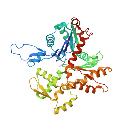
    Muscle contraction relies on the interaction of myosin motors with F-actin, which is regulated through a translocation of tropomyosin by the troponin complex in response to Ca 2+ The current model of muscle regulation holds that at relaxing (low-Ca 2+ ) conditions tropomyosin blocks myosin binding sites on F-actin, whereas at activating (high-Ca 2+ ) conditions tropomyosin translocation only partially exposes myosin binding sites on F-actin so that binding of rigor myosin is required to fully activate the thin filament (TF). Here we used a single-particle approach to helical reconstruction of frozen hydrated native cardiac TFs under relaxing and activating conditions to reveal the azimuthal movement of the tropomyosin on the surface of the native cardiac TF upon Ca 2+ activation. We demonstrate that at either relaxing or activating conditions tropomyosin is not constrained in one structural state, but rather is distributed between three structural positions on the surface of the TF. We show that two of these tropomyosin positions restrain actomyosin interactions, whereas in the third position, which is significantly enhanced at high Ca 2+ , tropomyosin does not block myosin binding sites on F-actin. Our data provide a structural framework for the enhanced activation of the cardiac TF over the skeletal TF by Ca 2+ and lead to a mechanistic model for the regulation of the cardiac TF.


  • Organizational Affiliation

    Department of Physiological Sciences, Eastern Virginia Medical School, Norfolk, VA 23507.


Macromolecules
Find similar proteins by:  (by identity cutoff)  |  3D Structure
Entity ID: 1
MoleculeChains Sequence LengthOrganismDetailsImage
Actin, alpha skeletal muscle
A, B, C, D, E
367Sus scrofaMutation(s): 0 
Gene Names: ACTA1ACTA
UniProt
Find proteins for P68137 (Sus scrofa)
Explore P68137 
Go to UniProtKB:  P68137
Entity Groups  
Sequence Clusters30% Identity50% Identity70% Identity90% Identity95% Identity100% Identity
UniProt GroupP68137
Sequence Annotations
Expand
  • Reference Sequence
Find similar proteins by:  (by identity cutoff)  |  3D Structure
Entity ID: 2
MoleculeChains Sequence LengthOrganismDetailsImage
cardiac alpha tropomyosinF,
G [auth H]
136Sus scrofaMutation(s): 0 
Sequence Annotations
Expand
  • Reference Sequence
Small Molecules
Ligands 2 Unique
IDChains Name / Formula / InChI Key2D Diagram3D Interactions
ADP
Query on ADP

Download Ideal Coordinates CCD File 
H [auth A],
J [auth B],
L [auth C],
N [auth D],
P [auth E]
ADENOSINE-5'-DIPHOSPHATE
C10 H15 N5 O10 P2
XTWYTFMLZFPYCI-KQYNXXCUSA-N
MG
Query on MG

Download Ideal Coordinates CCD File 
I [auth A],
K [auth B],
M [auth C],
O [auth D],
Q [auth E]
MAGNESIUM ION
Mg
JLVVSXFLKOJNIY-UHFFFAOYSA-N
Modified Residues  1 Unique
IDChains TypeFormula2D DiagramParent
HIC
Query on HIC
A, B, C, D, E
L-PEPTIDE LINKINGC7 H11 N3 O2HIS
Experimental Data & Validation

Experimental Data

  • Method: ELECTRON MICROSCOPY
  • Resolution: 11.0 Å
  • Aggregation State: HELICAL ARRAY 
  • Reconstruction Method: HELICAL 
EM Software:
TaskSoftware PackageVersion
RECONSTRUCTIONSPIDER

Structure Validation

View Full Validation Report


Entry History & Funding Information

Deposition Data


Funding OrganizationLocationGrant Number
AHAUnited StatesAid 560851

Revision History  (Full details and data files)

  • Version 1.0: 2017-08-02
    Type: Initial release
  • Version 1.1: 2017-08-09
    Changes: Database references