Combining EPR spectroscopy and X-ray crystallography to elucidate the structure and dynamics of conformationally constrained spin labels in T4 lysozyme single crystals.
Consentius, P., Gohlke, U., Loll, B., Alings, C., Heinemann, U., Wahl, M.C., Risse, T.(2017) Phys Chem Chem Phys 19: 20723-20734
- PubMed: 28740983
- DOI: https://doi.org/10.1039/c7cp03144k
- Primary Citation of Related Structures:
5NX0 - PubMed Abstract:
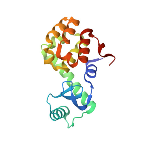
Electron paramagnetic resonance (EPR) spectroscopy in combination with site-directed spin labeling is used to investigate the structure and dynamics of conformationally constrained spin labels in T4 lysozyme single crystals. Within a single crystal, the oriented ensemble of spin bearing moieties results in a strong angle dependence of the EPR spectra. A quantitative description of the EPR spectra requires the determination of the unit cell orientation with respect to the sample tube and the orientation of the spin bearing moieties within the crystal lattice. Angle dependent EPR spectra were analyzed by line shape simulations using the stochastic Liouville equation approach developed by Freed and co-workers and an effective Hamiltonian approach. The gain in spectral information obtained from the EPR spectra of single crystalline samples taken at different frequencies, namely the X-band and Q-band, allows us to discriminate between motional models describing the spectra of isotropic solutions similarly well. In addition, it is shown that the angle dependent single crystal spectra allow us to identify two spin label rotamers with very similar side chain dynamics. These results demonstrate the utility of single crystal EPR spectroscopy in combination with spectral line shape simulation techniques to extract valuable dynamic information not readily available from the analysis of isotropic systems. In addition, it will be shown that the loss of electron density in high resolution diffraction experiments at room temperature does not allow us to conclude that there is significant structural disorder in the system.
Organizational Affiliation:
Freie Universität Berlin, Institute of Chemistry and Biochemistry, Takustr. 3, 14195 Berlin, Germany. risse@chemie.fu-berlin.de.