Multi-wavelength anomalous diffraction de novo phasing using a two-colour X-ray free-electron laser with wide tunability.
Gorel, A., Motomura, K., Fukuzawa, H., Doak, R.B., Grunbein, M.L., Hilpert, M., Inoue, I., Kloos, M., Kovacsova, G., Nango, E., Nass, K., Roome, C.M., Shoeman, R.L., Tanaka, R., Tono, K., Joti, Y., Yabashi, M., Iwata, S., Foucar, L., Ueda, K., Barends, T.R.M., Schlichting, I.(2017) Nat Commun 8: 1170-1170
- PubMed: 29079797
- DOI: https://doi.org/10.1038/s41467-017-00754-7
- Primary Citation of Related Structures:
5OER - PubMed Abstract:
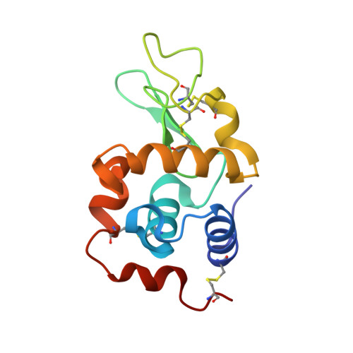
Serial femtosecond crystallography at X-ray free-electron lasers (XFELs) offers unprecedented possibilities for macromolecular structure determination of systems prone to radiation damage. However, de novo structure determination, i.e., without prior structural knowledge, is complicated by the inherent inaccuracy of serial femtosecond crystallography data. By its very nature, serial femtosecond crystallography data collection entails shot-to-shot fluctuations in X-ray wavelength and intensity as well as variations in crystal size and quality that must be averaged out. Hence, to obtain accurate diffraction intensities for de novo phasing, large numbers of diffraction patterns are required, and, concomitantly large volumes of sample and long X-ray free-electron laser beamtimes. Here we show that serial femtosecond crystallography data collected using simultaneous two-colour X-ray free-electron laser pulses can be used for multiple wavelength anomalous dispersion phasing. The phase angle determination is significantly more accurate than for single-colour phasing. We anticipate that two-colour multiple wavelength anomalous dispersion phasing will enhance structure determination of difficult-to-phase proteins at X-ray free-electron lasers.
Organizational Affiliation:
Max-Planck-Institut für medizinische Forschung, Jahnstrasse 29, Heidelberg, 69120, Germany.