5ONG

X-Ray crystal structure of a nucleosome core particle with its DNA site-specifically crosslinked to the histone octamer


Experimental Data Snapshot

  • Method: X-RAY DIFFRACTION
  • Resolution: 2.80 Å
  • R-Value Free: 0.247 
  • R-Value Work: 0.206 
  • R-Value Observed: 0.210 

wwPDB Validation   3D Report Full Report


This is version 1.2 of the entry. See complete history


Literature

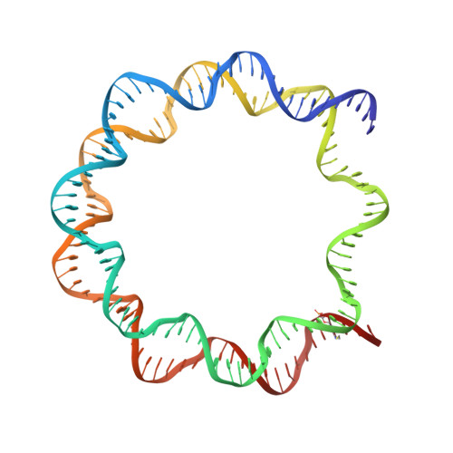
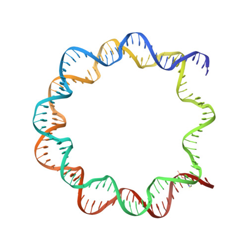
Site-Specific Disulfide Crosslinked Nucleosomes with Enhanced Stability.

Frouws, T.D.Barth, P.D.Richmond, T.J.

(2018) J Mol Biol 430: 45-57

  • DOI: https://doi.org/10.1016/j.jmb.2017.10.029
  • Primary Citation of Related Structures:  
    5OMX, 5ONG, 5ONW

  • PubMed Abstract: 

    We engineered nucleosome core particles (NCPs) with two site-specific cysteine crosslinks that increase the stability of the particle. The first disulfide was introduced between the two copies of H2A via an H2A-N38C point mutation, effectively crosslinking the two H2A/H2B heterodimers together to stabilize the histone octamer against H2A/H2B dimer dissociation. The second crosslink was engineered between an R40C point mutation on the N-terminal tail of H3 and the NCP DNA ends by the introduction of a convertible nucleotide. This crosslink maintains the nucleosome DNA in a fixed translational setting relative to the histone octamer and prevents dilution-driven dissociation. The X-ray crystal structures of NCPs containing the disulfides in isolation and in combination were determined. Both disulfides stabilize the structure of the NCP without disturbing the overall structure. Nucleosomes containing these modifications will be advantageous for biochemical and structural studies as a consequence of their greater resistance to dissociation during high dilution in purification, elevated salt for crystallization and vitrification for cryogenic electron microscopy.


  • Organizational Affiliation

    ETH Zürich, Institute of Molecular Biology and Biophysics, Otto-Stern-Weg 5, 8093 Zürich, Switzerland.


Macromolecules

Find similar proteins by:  (by identity cutoff)  |  3D Structure
Entity ID: 3
MoleculeChains Sequence LengthOrganismDetailsImage
Histone H3.2C [auth A],
G [auth E]
135Xenopus laevisMutation(s): 2 
UniProt
Find proteins for P84233 (Xenopus laevis)
Explore P84233 
Go to UniProtKB:  P84233
Entity Groups  
Sequence Clusters30% Identity50% Identity70% Identity90% Identity95% Identity100% Identity
UniProt GroupP84233
Sequence Annotations
Expand
  • Reference Sequence
Find similar proteins by:  (by identity cutoff)  |  3D Structure
Entity ID: 4
MoleculeChains Sequence LengthOrganismDetailsImage
Histone H4D [auth B],
H [auth F]
103Xenopus laevisMutation(s): 0 
UniProt
Find proteins for P62799 (Xenopus laevis)
Explore P62799 
Go to UniProtKB:  P62799
Entity Groups  
Sequence Clusters30% Identity50% Identity70% Identity90% Identity95% Identity100% Identity
UniProt GroupP62799
Sequence Annotations
Expand
  • Reference Sequence
Find similar proteins by:  (by identity cutoff)  |  3D Structure
Entity ID: 5
MoleculeChains Sequence LengthOrganismDetailsImage
Histone H2AE [auth C],
I [auth G]
129Xenopus laevisMutation(s): 0 
Gene Names: hist1h2ajLOC494591
UniProt
Find proteins for P06897 (Xenopus laevis)
Explore P06897 
Go to UniProtKB:  P06897
Entity Groups  
Sequence Clusters30% Identity50% Identity70% Identity90% Identity95% Identity100% Identity
UniProt GroupP06897
Sequence Annotations
Expand
  • Reference Sequence
Find similar proteins by:  (by identity cutoff)  |  3D Structure
Entity ID: 6
MoleculeChains Sequence LengthOrganismDetailsImage
Histone H2B 1.1F [auth D],
J [auth H]
122Xenopus laevisMutation(s): 0 
UniProt
Find proteins for P02281 (Xenopus laevis)
Explore P02281 
Go to UniProtKB:  P02281
Entity Groups  
Sequence Clusters30% Identity50% Identity70% Identity90% Identity95% Identity100% Identity
UniProt GroupP02281
Sequence Annotations
Expand
  • Reference Sequence
Find similar nucleic acids by:  (by identity cutoff)  |  3D Structure
Entity ID: 1
MoleculeChains LengthOrganismImage
DNA (147-MER)A [auth I]147Homo sapiens
Sequence Annotations
Expand
  • Reference Sequence
Find similar nucleic acids by:  (by identity cutoff)  |  3D Structure
Entity ID: 2
MoleculeChains LengthOrganismImage
DNA (147-MER)B [auth J]147Homo sapiens
Sequence Annotations
Expand
  • Reference Sequence
Small Molecules
Ligands 2 Unique
IDChains Name / Formula / InChI Key2D Diagram3D Interactions
MN
Query on MN

Download Ideal Coordinates CCD File 
AA [auth J]
BA [auth J]
CA [auth J]
FA [auth E]
K [auth I]
AA [auth J],
BA [auth J],
CA [auth J],
FA [auth E],
K [auth I],
L [auth I],
M [auth I],
N [auth I],
O [auth I],
P [auth I],
Q [auth I],
R [auth I],
S [auth I],
T [auth I],
U [auth J],
V [auth J],
W [auth J],
X [auth J],
Y [auth J],
Z [auth J]
MANGANESE (II) ION
Mn
WAEMQWOKJMHJLA-UHFFFAOYSA-N
CL
Query on CL

Download Ideal Coordinates CCD File 
DA [auth A],
EA [auth D],
GA [auth E],
HA [auth G]
CHLORIDE ION
Cl
VEXZGXHMUGYJMC-UHFFFAOYSA-M
Experimental Data & Validation

Experimental Data

  • Method: X-RAY DIFFRACTION
  • Resolution: 2.80 Å
  • R-Value Free: 0.247 
  • R-Value Work: 0.206 
  • R-Value Observed: 0.210 
  • Space Group: P 21 21 21
Unit Cell:
Length ( Å )Angle ( ˚ )
a = 106.24α = 90
b = 183.13β = 90
c = 109.49γ = 90
Software Package:
Software NamePurpose
PHENIXrefinement
XDSdata reduction
SCALAdata scaling
PHASERphasing

Structure Validation

View Full Validation Report


Entry History & Funding Information

Deposition Data


Funding OrganizationLocationGrant Number
Louis Jeantet PrizeSwitzerland--

Revision History  (Full details and data files)

  • Version 1.0: 2017-11-22
    Type: Initial release
  • Version 1.1: 2017-12-27
    Changes: Database references
  • Version 1.2: 2024-01-17
    Changes: Data collection, Database references, Derived calculations, Refinement description