5THR

Cryo-EM structure of a BG505 Env-sCD4-17b-8ANC195 complex


Experimental Data Snapshot

  • Method: ELECTRON MICROSCOPY
  • Resolution: 8.90 Å
  • Aggregation State: PARTICLE 
  • Reconstruction Method: SINGLE PARTICLE 

wwPDB Validation   3D Report Full Report


This is version 2.0 of the entry. See complete history


Literature

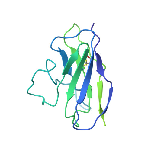
Cryo-EM structure of a CD4-bound open HIV-1 envelope trimer reveals structural rearrangements of the gp120 V1V2 loop.

Wang, H.Cohen, A.A.Galimidi, R.P.Gristick, H.B.Jensen, G.J.Bjorkman, P.J.

(2016) Proc Natl Acad Sci U S A 113: E7151-E7158

  • DOI: https://doi.org/10.1073/pnas.1615939113
  • Primary Citation of Related Structures:  
    5THR

  • PubMed Abstract: 

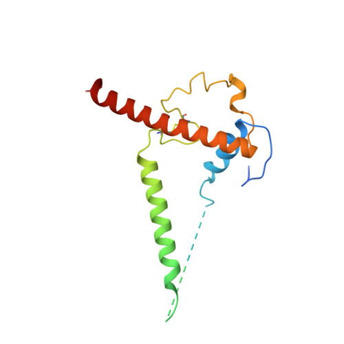
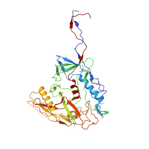
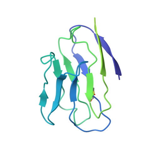
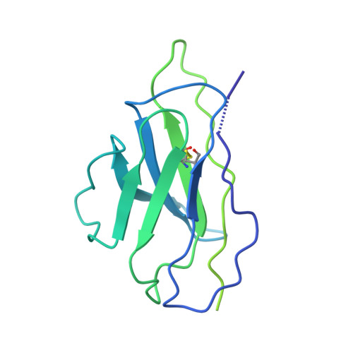
    The HIV-1 envelope (Env) glycoprotein, a trimer of gp120-gp41 heterodimers, relies on conformational flexibility to function in fusing the viral and host membranes. Fusion is achieved after gp120 binds to CD4, the HIV-1 receptor, and a coreceptor, capturing an open conformational state in which the fusion machinery on gp41 gains access to the target cell membrane. In the well-characterized closed Env conformation, the gp120 V1V2 loops interact at the apex of the Env trimer. Less is known about the structure of the open CD4-bound state, in which the V1V2 loops must rearrange and separate to allow access to the coreceptor binding site. We identified two anti-HIV-1 antibodies, the coreceptor mimicking antibody 17b and the gp120-gp41 interface-spanning antibody 8ANC195, that can be added as Fabs to a soluble native-like Env trimer to stabilize it in a CD4-bound conformation. Here, we present an 8.9-Å cryo-electron microscopy structure of a BG505 Env-sCD4-17b-8ANC195 complex, which reveals large structural rearrangements in gp120, but small changes in gp41, compared with closed Env structures. The gp120 protomers are rotated and separated in the CD4-bound structure, and the three V1V2 loops are displaced by ∼40 Å from their positions at the trimer apex in closed Env to the sides of the trimer in positions adjacent to, and interacting with, the three bound CD4s. These results are relevant to understanding CD4-induced conformational changes leading to coreceptor binding and fusion, and HIV-1 Env conformational dynamics, and describe a target structure relevant to drug design and vaccine efforts.


  • Organizational Affiliation

    Division of Biology and Biological Engineering, California Institute of Technology, Pasadena, CA 91125.


Macromolecules
Find similar proteins by:  (by identity cutoff)  |  3D Structure
Entity ID: 1
MoleculeChains Sequence LengthOrganismDetailsImage
BG505 SOSIP gp41
A, B, C
153Human immunodeficiency virus 1Mutation(s): 0 
Gene Names: env
UniProt
Find proteins for Q2N0S6 (Human immunodeficiency virus 1)
Explore Q2N0S6 
Go to UniProtKB:  Q2N0S6
Entity Groups  
Sequence Clusters30% Identity50% Identity70% Identity90% Identity95% Identity100% Identity
UniProt GroupQ2N0S6
Sequence Annotations
Expand
  • Reference Sequence
Find similar proteins by:  (by identity cutoff)  |  3D Structure
Entity ID: 2
MoleculeChains Sequence LengthOrganismDetailsImage
BG505 SOSIP gp120
D, E, F
481Human immunodeficiency virus 1Mutation(s): 0 
Gene Names: env
UniProt
Find proteins for Q2N0S6 (Human immunodeficiency virus 1)
Explore Q2N0S6 
Go to UniProtKB:  Q2N0S6
Entity Groups  
Sequence Clusters30% Identity50% Identity70% Identity90% Identity95% Identity100% Identity
UniProt GroupQ2N0S6
Sequence Annotations
Expand
  • Reference Sequence
Find similar proteins by:  (by identity cutoff)  |  3D Structure
Entity ID: 3
MoleculeChains Sequence LengthOrganismDetailsImage
T-cell surface glycoprotein CD4
G, H, I
192Homo sapiensMutation(s): 0 
Gene Names: CD4
UniProt & NIH Common Fund Data Resources
Find proteins for P01730 (Homo sapiens)
Explore P01730 
Go to UniProtKB:  P01730
PHAROS:  P01730
GTEx:  ENSG00000010610 
Entity Groups  
Sequence Clusters30% Identity50% Identity70% Identity90% Identity95% Identity100% Identity
UniProt GroupP01730
Sequence Annotations
Expand
  • Reference Sequence
Find similar proteins by:  (by identity cutoff)  |  3D Structure
Entity ID: 4
MoleculeChains Sequence LengthOrganismDetailsImage
17b Fab VL domain
J, K, L
214Homo sapiensMutation(s): 0 
Entity Groups  
Sequence Clusters30% Identity50% Identity70% Identity90% Identity95% Identity100% Identity
Sequence Annotations
Expand
  • Reference Sequence
Find similar proteins by:  (by identity cutoff)  |  3D Structure
Entity ID: 5
MoleculeChains Sequence LengthOrganismDetailsImage
17b Fab VH domain
M, N, O
229Homo sapiensMutation(s): 0 
Entity Groups  
Sequence Clusters30% Identity50% Identity70% Identity90% Identity95% Identity100% Identity
Sequence Annotations
Expand
  • Reference Sequence
Find similar proteins by:  (by identity cutoff)  |  3D Structure
Entity ID: 6
MoleculeChains Sequence LengthOrganismDetailsImage
8ANC195 G52K5 VH domain
P, R, T
244Homo sapiensMutation(s): 0 
Entity Groups  
Sequence Clusters30% Identity50% Identity70% Identity90% Identity95% Identity100% Identity
Sequence Annotations
Expand
  • Reference Sequence
Find similar proteins by:  (by identity cutoff)  |  3D Structure
Entity ID: 7
MoleculeChains Sequence LengthOrganismDetailsImage
8ANC195 G52K5 VL domain
Q, S, U
215Homo sapiensMutation(s): 0 
Entity Groups  
Sequence Clusters30% Identity50% Identity70% Identity90% Identity95% Identity100% Identity
Sequence Annotations
Expand
  • Reference Sequence
Oligosaccharides

Help

Entity ID: 8
MoleculeChains Length2D Diagram Glycosylation3D Interactions
alpha-D-mannopyranose-(1-3)-beta-D-mannopyranose-(1-4)-2-acetamido-2-deoxy-beta-D-glucopyranose-(1-4)-2-acetamido-2-deoxy-beta-D-glucopyranoseBA [auth b],
V,
Y
4N-Glycosylation
Glycosylation Resources
GlyTouCan:  G81315DD
GlyCosmos:  G81315DD
GlyGen:  G81315DD
Entity ID: 9
MoleculeChains Length2D Diagram Glycosylation3D Interactions
alpha-D-mannopyranose-(1-3)-[alpha-D-mannopyranose-(1-6)]beta-D-mannopyranose-(1-4)-2-acetamido-2-deoxy-beta-D-glucopyranose-(1-4)-2-acetamido-2-deoxy-beta-D-glucopyranose
AA [auth a],
CA [auth c],
DA [auth d],
W,
X,
AA [auth a],
CA [auth c],
DA [auth d],
W,
X,
Z
5N-Glycosylation
Glycosylation Resources
GlyTouCan:  G22768VO
GlyCosmos:  G22768VO
GlyGen:  G22768VO
Experimental Data & Validation

Experimental Data

  • Method: ELECTRON MICROSCOPY
  • Resolution: 8.90 Å
  • Aggregation State: PARTICLE 
  • Reconstruction Method: SINGLE PARTICLE 
EM Software:
TaskSoftware PackageVersion
RECONSTRUCTIONRELION
MODEL REFINEMENTPHENIX

Structure Validation

View Full Validation Report


Entry History & Funding Information

Deposition Data


Funding OrganizationLocationGrant Number
National Institutes of Health/National Institute of General Medical Sciences (NIH/NIGMS)United States2 P50 GM082545-06
National Institutes of Health/National Institute Of Allergy and Infectious Diseases (NIH/NIAID)United StatesHIVRAD P01 AI100148
Bill & Melinda Gates FoundationUnited StatesCollaboration for AIDS Vaccine Discovery Grant 1040753

Revision History  (Full details and data files)

  • Version 1.0: 2016-11-16
    Type: Initial release
  • Version 1.1: 2016-11-30
    Changes: Database references
  • Version 1.2: 2017-09-27
    Changes: Author supporting evidence, Data collection, Database references
  • Version 1.3: 2018-07-18
    Changes: Data collection
  • Version 1.4: 2018-10-03
    Changes: Data collection, Other, Refinement description, Structure summary
  • Version 1.5: 2019-12-11
    Changes: Author supporting evidence
  • Version 1.6: 2019-12-18
    Changes: Other
  • Version 2.0: 2020-07-29
    Type: Remediation
    Reason: Carbohydrate remediation
    Changes: Advisory, Atomic model, Data collection, Derived calculations, Structure summary