Structural Consequences of Calmodulin EF Hand Mutations.
Piazza, M., Taiakina, V., Dieckmann, T., Guillemette, J.G.(2017) Biochemistry 56: 944-956
- PubMed: 28121131
- DOI: https://doi.org/10.1021/acs.biochem.6b01296
- Primary Citation of Related Structures:
5TP5, 5TP6 - PubMed Abstract:
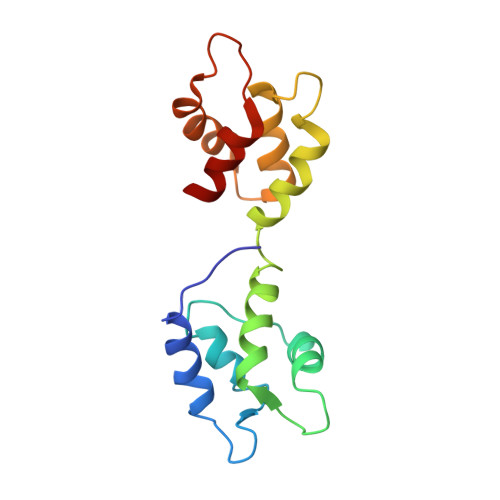
Calmodulin (CaM) is a cytosolic Ca 2+ -binding protein that serves as a control element for many enzymes. It consists of two globular domains, each containing two EF hand pairs capable of binding Ca 2+ , joined by a flexible central linker region. CaM is able to bind and activate its target proteins in the Ca 2+ -replete and Ca 2+ -deplete forms. To study the Ca 2+ -dependent/independent properties of binding and activation of target proteins by CaM, CaM constructs with Ca 2+ -binding disrupting mutations of Asp to Ala at position one of each EF hand have been used. These CaM mutant proteins are deficient in binding Ca 2+ in either the N-lobe EF hands (CaM 12 ), C-lobe EF hands (CaM 34 ), or all four EF hands (CaM 1234 ). To investigate potential structural changes these mutations may cause, we performed detailed NMR studies of CaM 12 , CaM 34 , and CaM 1234 including determining the solution structure of CaM 1234 . We then investigated if these CaM mutants affected the interaction of CaM with a target protein known to interact with apoCaM by determining the solution structure of CaM 34 bound to the iNOS CaM binding domain peptide. The structures provide direct structural evidence of changes that are present in these Ca 2+ -deficient CaM mutants and show these mutations increase the hydrophobic exposed surface and decrease the electronegative surface potential throughout each lobe of CaM. These Ca 2+ -deficient CaM mutants may not be a true representation of apoCaM and may not allow for native-like interactions of apoCaM with its target proteins.
Organizational Affiliation:
Department of Chemistry, University of Waterloo , Waterloo, Ontario N2L 3G1, Canada.