5TY8

Identification of a New Zinc Binding Chemotype by Fragment Screening


Experimental Data Snapshot

  • Method: X-RAY DIFFRACTION
  • Resolution: 1.45 Å
  • R-Value Free: 0.163 
  • R-Value Work: 0.121 
  • R-Value Observed: 0.123 

wwPDB Validation   3D Report Full Report


Ligand Structure Quality Assessment 


This is version 1.4 of the entry. See complete history


Literature

Identification of a New Zinc Binding Chemotype by Fragment Screening.

Chrysanthopoulos, P.K.Mujumdar, P.Woods, L.A.Dolezal, O.Ren, B.Peat, T.S.Poulsen, S.A.

(2017) J Med Chem 60: 7333-7349

  • DOI: https://doi.org/10.1021/acs.jmedchem.7b00606
  • Primary Citation of Related Structures:  
    5TXY, 5TY1, 5TY8, 5TY9, 5TYA, 5U0D, 5U0E, 5U0F, 5U0G, 5VGY

  • PubMed Abstract: 

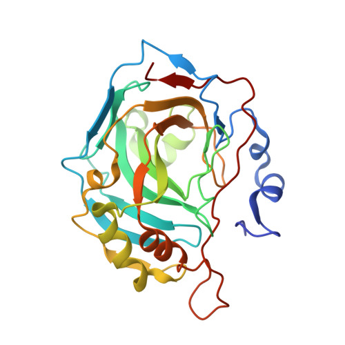
    The discovery of a new zinc binding chemotype from screening a nonbiased fragment library is reported. Using the orthogonal fragment screening methods of native state mass spectrometry and surface plasmon resonance a 3-unsubstituted 2,4-oxazolidinedione fragment was found to have low micromolar binding affinity to the zinc metalloenzyme carbonic anhydrase II (CA II). This affinity approached that of fragment sized primary benzenesulfonamides, the classical zinc binding group found in most CA II inhibitors. Protein X-ray crystallography established that 3-unsubstituted 2,4-oxazolidinediones bound to CA II via an interaction of the acidic ring nitrogen with the CA II active site zinc, as well as two hydrogen bonds between the oxazolidinedione ring oxygen and the CA II protein backbone. Furthermore, 3-unsubstituted 2,4-oxazolidinediones appear to be a viable starting point for the development of an alternative class of CA inhibitor, wherein the medicinal chemistry pedigree of primary sulfonamides has dominated for several decades.


  • Organizational Affiliation

    Griffith University , Griffith Institute for Drug Discovery, Nathan, Brisbane, Queensland 4111, Australia.


Macromolecules
Find similar proteins by:  (by identity cutoff)  |  3D Structure
Entity ID: 1
MoleculeChains Sequence LengthOrganismDetailsImage
Carbonic anhydrase 2260Homo sapiensMutation(s): 0 
Gene Names: CA2
EC: 4.2.1.1
UniProt & NIH Common Fund Data Resources
Find proteins for P00918 (Homo sapiens)
Explore P00918 
Go to UniProtKB:  P00918
PHAROS:  P00918
GTEx:  ENSG00000104267 
Entity Groups  
Sequence Clusters30% Identity50% Identity70% Identity90% Identity95% Identity100% Identity
UniProt GroupP00918
Sequence Annotations
Expand
  • Reference Sequence
Small Molecules
Ligands 2 Unique
IDChains Name / Formula / InChI Key2D Diagram3D Interactions
7Q1
Query on 7Q1

Download Ideal Coordinates CCD File 
C [auth A](5R)-5-phenyl-1,3-oxazolidine-2,4-dione
C9 H7 N O3
SBYYYVAMWBVIIX-SSDOTTSWSA-N
ZN
Query on ZN

Download Ideal Coordinates CCD File 
B [auth A]ZINC ION
Zn
PTFCDOFLOPIGGS-UHFFFAOYSA-N
Binding Affinity Annotations 
IDSourceBinding Affinity
7Q1 BindingDB:  5TY8 Kd: min: 2600, max: 3500 (nM) from 3 assay(s)
Binding MOAD:  5TY8 Kd: 3500 (nM) from 1 assay(s)
Experimental Data & Validation

Experimental Data

  • Method: X-RAY DIFFRACTION
  • Resolution: 1.45 Å
  • R-Value Free: 0.163 
  • R-Value Work: 0.121 
  • R-Value Observed: 0.123 
  • Space Group: P 1 21 1
Unit Cell:
Length ( Å )Angle ( ˚ )
a = 42.522α = 90
b = 41.427β = 104.47
c = 72.138γ = 90
Software Package:
Software NamePurpose
REFMACrefinement
XDSdata reduction
Aimlessdata scaling
PHASERphasing

Structure Validation

View Full Validation Report


Ligand Structure Quality Assessment 


Entry History & Funding Information

Deposition Data


Funding OrganizationLocationGrant Number
Australian Research Council (ARC)Australia--

Revision History  (Full details and data files)

  • Version 1.0: 2017-08-30
    Type: Initial release
  • Version 1.1: 2017-09-27
    Changes: Database references
  • Version 1.2: 2018-01-17
    Changes: Author supporting evidence
  • Version 1.3: 2020-01-01
    Changes: Author supporting evidence
  • Version 1.4: 2023-10-04
    Changes: Data collection, Database references, Refinement description