5UBO

Mical-oxidized Actin complex with Gelsolin Segment 1


Experimental Data Snapshot

  • Method: X-RAY DIFFRACTION
  • Resolution: 2.39 Å
  • R-Value Free: 0.191 
  • R-Value Work: 0.172 
  • R-Value Observed: 0.173 

wwPDB Validation   3D Report Full Report


Ligand Structure Quality Assessment 


This is version 1.3 of the entry. See complete history


Literature

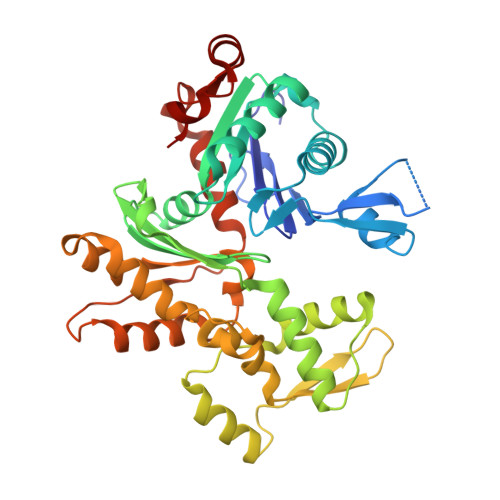
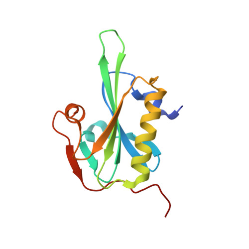
Catastrophic disassembly of actin filaments via Mical-mediated oxidation.

Grintsevich, E.E.Ge, P.Sawaya, M.R.Yesilyurt, H.G.Terman, J.R.Zhou, Z.H.Reisler, E.

(2017) Nat Commun 8: 2183-2183

  • DOI: https://doi.org/10.1038/s41467-017-02357-8
  • Primary Citation of Related Structures:  
    5UBO, 6AV9, 6AVB

  • PubMed Abstract: 

    Actin filament assembly and disassembly are vital for cell functions. MICAL Redox enzymes are important post-translational effectors of actin that stereo-specifically oxidize actin's M44 and M47 residues to induce cellular F-actin disassembly. Here we show that Mical-oxidized (Mox) actin can undergo extremely fast (84 subunits/s) disassembly, which depends on F-actin's nucleotide-bound state. Using near-atomic resolution cryoEM reconstruction and single filament TIRF microscopy we identify two dynamic and structural states of Mox-actin. Modeling actin's D-loop region based on our 3.9 Å cryoEM reconstruction suggests that oxidation by Mical reorients the side chain of M44 and induces a new intermolecular interaction of actin residue M47 (M47-O-T351). Site-directed mutagenesis reveals that this interaction promotes Mox-actin instability. Moreover, we find that Mical oxidation of actin allows for cofilin-mediated severing even in the presence of inorganic phosphate. Thus, in conjunction with cofilin, Mical oxidation of actin promotes F-actin disassembly independent of the nucleotide-bound state.


  • Organizational Affiliation

    Department of Chemistry and Biochemistry, University of California (UCLA), Los Angeles, CA, 90095, USA.


Macromolecules
Find similar proteins by:  (by identity cutoff)  |  3D Structure
Entity ID: 1
MoleculeChains Sequence LengthOrganismDetailsImage
Actin, alpha skeletal muscle377Oryctolagus cuniculusMutation(s): 0 
UniProt
Find proteins for P68135 (Oryctolagus cuniculus)
Explore P68135 
Go to UniProtKB:  P68135
Entity Groups  
Sequence Clusters30% Identity50% Identity70% Identity90% Identity95% Identity100% Identity
UniProt GroupP68135
Sequence Annotations
Expand
  • Reference Sequence
Find similar proteins by:  (by identity cutoff)  |  3D Structure
Entity ID: 2
MoleculeChains Sequence LengthOrganismDetailsImage
GelsolinB [auth S]127Homo sapiensMutation(s): 0 
Gene Names: GSN
UniProt & NIH Common Fund Data Resources
Find proteins for P06396 (Homo sapiens)
Explore P06396 
Go to UniProtKB:  P06396
PHAROS:  P06396
GTEx:  ENSG00000148180 
Entity Groups  
Sequence Clusters30% Identity50% Identity70% Identity90% Identity95% Identity100% Identity
UniProt GroupP06396
Sequence Annotations
Expand
  • Reference Sequence
Experimental Data & Validation

Experimental Data

  • Method: X-RAY DIFFRACTION
  • Resolution: 2.39 Å
  • R-Value Free: 0.191 
  • R-Value Work: 0.172 
  • R-Value Observed: 0.173 
  • Space Group: P 21 21 21
Unit Cell:
Length ( Å )Angle ( ˚ )
a = 56.2α = 90
b = 69.25β = 90
c = 179.18γ = 90
Software Package:
Software NamePurpose
BUSTERrefinement
XSCALEdata scaling
PDB_EXTRACTdata extraction
XDSdata reduction
Cootmodel building
XDSdata processing
MOSFLMdata reduction
X-PLORmodel building
CCP4data scaling
X-PLORphasing

Structure Validation

View Full Validation Report


Ligand Structure Quality Assessment 


Entry History 

Deposition Data

Revision History  (Full details and data files)

  • Version 1.0: 2017-12-20
    Type: Initial release
  • Version 1.1: 2018-01-17
    Changes: Author supporting evidence
  • Version 1.2: 2019-10-23
    Changes: Data collection, Database references
  • Version 1.3: 2023-10-04
    Changes: Data collection, Database references, Derived calculations, Refinement description