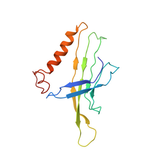
5VLY

Asymmetric unit for the coat proteins of phage Qbeta

  • Classification: VIRUS
  • Organism(s): Qubevirus durum
  • Mutation(s): No 

  • Deposited: 2017-04-26 Released: 2017-10-18 
  • Deposition Author(s): Cui, Z., Zhang, J.
  • Funding Organization(s): Welch Foundation, National Institutes of Health/National Institute of General Medical Sciences (NIH/NIGMS), Public Health Service

Experimental Data Snapshot

  • Method: ELECTRON MICROSCOPY
  • Resolution: 3.30 Å
  • Aggregation State: PARTICLE 
  • Reconstruction Method: SINGLE PARTICLE 

wwPDB Validation   3D Report Full Report


This is version 1.4 of the entry. See complete history


Literature

Structures of Q beta virions, virus-like particles, and the Q beta-MurA complex reveal internal coat proteins and the mechanism of host lysis.

Cui, Z.Gorzelnik, K.V.Chang, J.Y.Langlais, C.Jakana, J.Young, R.Zhang, J.

(2017) Proc Natl Acad Sci U S A 114: 11697-11702

  • DOI: https://doi.org/10.1073/pnas.1707102114
  • Primary Citation of Related Structures:  
    5VLY, 5VLZ, 5VM7

  • PubMed Abstract: 

    In single-stranded RNA bacteriophages (ssRNA phages) a single copy of the maturation protein binds the genomic RNA (gRNA) and is required for attachment of the phage to the host pilus. For the canonical Allolevivirus Qβ the maturation protein, A 2 , has an additional role as the lysis protein, by its ability to bind and inhibit MurA, which is involved in peptidoglycan biosynthesis. Here, we determined structures of Qβ virions, virus-like particles, and the Qβ-MurA complex using single-particle cryoelectron microscopy, at 4.7-Å, 3.3-Å, and 6.1-Å resolutions, respectively. We identified the outer surface of the β-region in A 2 as the MurA-binding interface. Moreover, the pattern of MurA mutations that block Qβ lysis and the conformational changes of MurA that facilitate A 2 binding were found to be due to the intimate fit between A 2 and the region encompassing the closed catalytic cleft of substrate-liganded MurA. Additionally, by comparing the Qβ virion with Qβ virus-like particles that lack a maturation protein, we observed a structural rearrangement in the capsid coat proteins that is required to package the viral gRNA in its dominant conformation. Unexpectedly, we found a coat protein dimer sequestered in the interior of the virion. This coat protein dimer binds to the gRNA and interacts with the buried α-region of A 2 , suggesting that it is sequestered during the early stage of capsid formation to promote the gRNA condensation required for genome packaging. These internalized coat proteins are the most asymmetrically arranged major capsid proteins yet observed in virus structures.


  • Organizational Affiliation

    Department of Biochemistry and Biophysics, Center for Phage Technology, Texas A&M University, College Station, TX 77843.


Macromolecules
Find similar proteins by:  (by identity cutoff)  |  3D Structure
Entity ID: 1
MoleculeChains Sequence LengthOrganismDetailsImage
Capsid protein
A, B, C
133Qubevirus durumMutation(s): 0 
UniProt
Find proteins for P03615 (Escherichia virus Qbeta)
Explore P03615 
Go to UniProtKB:  P03615
Entity Groups  
Sequence Clusters30% Identity50% Identity70% Identity90% Identity95% Identity100% Identity
UniProt GroupP03615
Sequence Annotations
Expand
  • Reference Sequence
Experimental Data & Validation

Experimental Data

  • Method: ELECTRON MICROSCOPY
  • Resolution: 3.30 Å
  • Aggregation State: PARTICLE 
  • Reconstruction Method: SINGLE PARTICLE 
EM Software:
TaskSoftware PackageVersion
RECONSTRUCTIONRELION1.4
MODEL REFINEMENTPHENIX

Structure Validation

View Full Validation Report


Entry History & Funding Information

Deposition Data


Funding OrganizationLocationGrant Number
Welch FoundationUnited StatesA-1863
National Institutes of Health/National Institute of General Medical Sciences (NIH/NIGMS)United StatesP41GM103832
National Institutes of Health/National Institute of General Medical Sciences (NIH/NIGMS)United StatesU24GM116787
Public Health ServiceUnited StatesGM27099

Revision History  (Full details and data files)

  • Version 1.0: 2017-10-18
    Type: Initial release
  • Version 1.1: 2018-04-18
    Changes: Data collection, Database references
  • Version 1.2: 2018-07-18
    Changes: Data collection, Experimental preparation
  • Version 1.3: 2020-01-01
    Changes: Author supporting evidence
  • Version 1.4: 2024-03-13
    Changes: Data collection, Database references, Derived calculations