5WK7

P450cam mutant R186A


Experimental Data Snapshot

  • Method: X-RAY DIFFRACTION
  • Resolution: 1.98 Å
  • R-Value Free: 0.215 
  • R-Value Work: 0.163 
  • R-Value Observed: 0.167 

wwPDB Validation   3D Report Full Report


Ligand Structure Quality Assessment 


This is version 1.4 of the entry. See complete history


Literature

Effect of Redox Partner Binding on Cytochrome P450 Conformational Dynamics.

Batabyal, D.Richards, L.S.Poulos, T.L.

(2017) J Am Chem Soc 139: 13193-13199

  • DOI: https://doi.org/10.1021/jacs.7b07656
  • Primary Citation of Related Structures:  
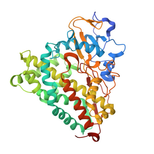
    5WK7, 5WK9

  • PubMed Abstract: 

    Previous crystal structures of cytochrome P450cam complexed with its redox partner, putidaredoxin (Pdx), shows that P450cam adopts the open conformation. It has been hypothesized that the Pdx-induced shift toward the open state frees the essential Asp251 from salt bridges with Arg186 and Lys178 so that Asp251 can participate in a proton relay network required for O 2 activation. This in part explains why P450cam has such a strict requirement for Pdx. One problem with this view is that looser substrate-protein interactions in the open state may not be compatible with the observed regio- and stereoselective hydroxylation. In the present study, molecular dynamics simulations show that Pdx binding favors a conformation that stabilizes the active site and decreases camphor mobility yet retains a partially open conformation compatible with the required proton relay network. The R186A mutant which frees Asp251 in the absence of Pdx retains good enzyme activity, and the crystal structure shows that product, 5-exo-hydroxycamphor, is bound. This indicates that rupture of the Asp251-Arg186 relaxes selectivity with respect to source of electrons and enables X-ray generated reducing equivalents to support substrate hydroxylation. These combined computational and experimental results are consistent with the proposed role of Pdx in assisting the release of Asp251 from ion pairs so that it can participate in proton-coupled electron transfer.


  • Organizational Affiliation

    Departments of Molecular Biology and Biochemistry, Pharmaceutical Sciences, and Chemistry, University of California , Irvine, California 92697-3900, United States.


Macromolecules
Find similar proteins by:  (by identity cutoff)  |  3D Structure
Entity ID: 1
MoleculeChains Sequence LengthOrganismDetailsImage
Camphor 5-monooxygenase415Pseudomonas putidaMutation(s): 2 
Gene Names: camCcyp101
EC: 1.14.15.1
UniProt
Find proteins for P00183 (Pseudomonas putida)
Explore P00183 
Go to UniProtKB:  P00183
Entity Groups  
Sequence Clusters30% Identity50% Identity70% Identity90% Identity95% Identity100% Identity
UniProt GroupP00183
Sequence Annotations
Expand
  • Reference Sequence
Experimental Data & Validation

Experimental Data

  • Method: X-RAY DIFFRACTION
  • Resolution: 1.98 Å
  • R-Value Free: 0.215 
  • R-Value Work: 0.163 
  • R-Value Observed: 0.167 
  • Space Group: P 1 21 1
Unit Cell:
Length ( Å )Angle ( ˚ )
a = 56.82α = 90
b = 58.42β = 104.97
c = 57.25γ = 90
Software Package:
Software NamePurpose
PHENIXrefinement
MOSFLMdata reduction
Aimlessdata scaling
MOLREPphasing

Structure Validation

View Full Validation Report


Ligand Structure Quality Assessment 


Entry History & Funding Information

Deposition Data


Funding OrganizationLocationGrant Number
National Institutes of Health/National Institute of General Medical Sciences (NIH/NIGMS)United StatesGM57353

Revision History  (Full details and data files)

  • Version 1.0: 2017-09-06
    Type: Initial release
  • Version 1.1: 2017-09-20
    Changes: Database references
  • Version 1.2: 2017-09-27
    Changes: Author supporting evidence, Database references
  • Version 1.3: 2020-01-01
    Changes: Author supporting evidence, Database references
  • Version 1.4: 2023-10-04
    Changes: Data collection, Database references, Refinement description