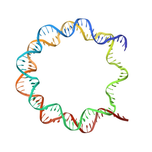
5XM1

The mouse nucleosome structure containing H2A, H2B type3-A, H3mm7, and H4


Experimental Data Snapshot

  • Method: X-RAY DIFFRACTION
  • Resolution: 3.45 Å
  • R-Value Free: 0.254 
  • R-Value Work: 0.200 
  • R-Value Observed: 0.204 

wwPDB Validation   3D Report Full Report


This is version 1.2 of the entry. See complete history


Literature

Histone H3.3 sub-variant H3mm7 is required for normal skeletal muscle regeneration.

Harada, A.Maehara, K.Ono, Y.Taguchi, H.Yoshioka, K.Kitajima, Y.Xie, Y.Sato, Y.Iwasaki, T.Nogami, J.Okada, S.Komatsu, T.Semba, Y.Takemoto, T.Kimura, H.Kurumizaka, H.Ohkawa, Y.

(2018) Nat Commun 9: 1400-1400

  • DOI: https://doi.org/10.1038/s41467-018-03845-1
  • Primary Citation of Related Structures:  
    5XM0, 5XM1

  • PubMed Abstract: 

    Regulation of gene expression requires selective incorporation of histone H3 variant H3.3 into chromatin. Histone H3.3 has several subsidiary variants but their functions are unclear. Here we characterize the function of histone H3.3 sub-variant, H3mm7, which is expressed in skeletal muscle satellite cells. H3mm7 knockout mice demonstrate an essential role of H3mm7 in skeletal muscle regeneration. Chromatin analysis reveals that H3mm7 facilitates transcription by forming an open chromatin structure around promoter regions including those of myogenic genes. The crystal structure of the nucleosome containing H3mm7 reveals that, unlike the S57 residue of other H3 proteins, the H3mm7-specific A57 residue cannot form a hydrogen bond with the R40 residue of the cognate H4 molecule. Consequently, the H3mm7 nucleosome is unstable in vitro and exhibited higher mobility in vivo compared with the H3.3 nucleosome. We conclude that the unstable H3mm7 nucleosome may be required for proper skeletal muscle differentiation.


  • Organizational Affiliation

    Division of Transcriptomics, Medical Institute of Bioregulation, Kyushu University, 3-1-1 Maidashi, Higashi, Fukuoka, 812-0054, Japan.


Macromolecules

Find similar proteins by:  (by identity cutoff)  |  3D Structure
Entity ID: 1
MoleculeChains Sequence LengthOrganismDetailsImage
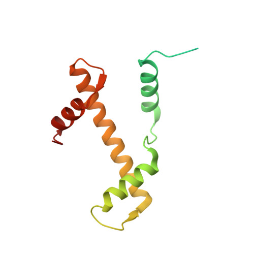
Histone H3mm7
A, E
139Mus musculusMutation(s): 0 
UniProt
Find proteins for P84244 (Mus musculus)
Explore P84244 
Go to UniProtKB:  P84244
Entity Groups  
Sequence Clusters30% Identity50% Identity70% Identity90% Identity95% Identity100% Identity
UniProt GroupP84244
Sequence Annotations
Expand
  • Reference Sequence
Find similar proteins by:  (by identity cutoff)  |  3D Structure
Entity ID: 2
MoleculeChains Sequence LengthOrganismDetailsImage
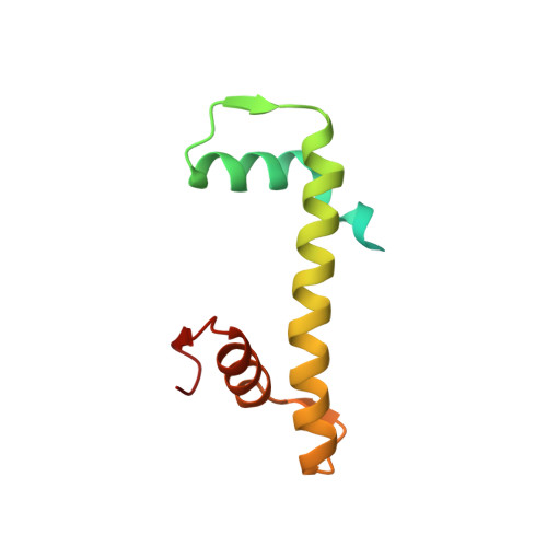
Histone H4
B, F
106Mus musculusMutation(s): 0 
Gene Names: 
UniProt & NIH Common Fund Data Resources
Find proteins for P62806 (Mus musculus)
Explore P62806 
Go to UniProtKB:  P62806
IMPC:  MGI:2448443
Entity Groups  
Sequence Clusters30% Identity50% Identity70% Identity90% Identity95% Identity100% Identity
UniProt GroupP62806
Sequence Annotations
Expand
  • Reference Sequence
Find similar proteins by:  (by identity cutoff)  |  3D Structure
Entity ID: 3
MoleculeChains Sequence LengthOrganismDetailsImage
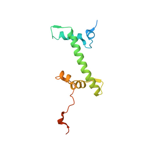
Histone H2A type 1-B
C, G
133Mus musculusMutation(s): 0 
Gene Names: Hist1h2ab
UniProt
Find proteins for C0HKE1 (Mus musculus)
Explore C0HKE1 
Go to UniProtKB:  C0HKE1
Entity Groups  
Sequence Clusters30% Identity50% Identity70% Identity90% Identity95% Identity100% Identity
UniProt GroupC0HKE1
Sequence Annotations
Expand
  • Reference Sequence
Find similar proteins by:  (by identity cutoff)  |  3D Structure
Entity ID: 4
MoleculeChains Sequence LengthOrganismDetailsImage
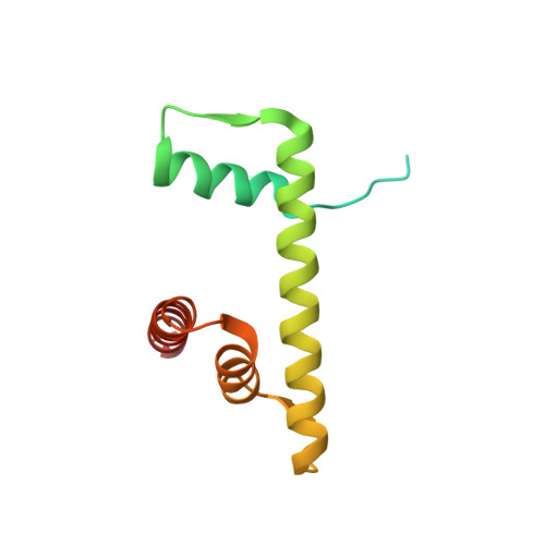
Histone H2B type 3-A
D, H
129Mus musculusMutation(s): 0 
Gene Names: Hist3h2ba
UniProt
Find proteins for Q9D2U9 (Mus musculus)
Explore Q9D2U9 
Go to UniProtKB:  Q9D2U9
Entity Groups  
Sequence Clusters30% Identity50% Identity70% Identity90% Identity95% Identity100% Identity
UniProt GroupQ9D2U9
Sequence Annotations
Expand
  • Reference Sequence
Find similar nucleic acids by:  (by identity cutoff)  |  3D Structure
Entity ID: 5
MoleculeChains LengthOrganismImage
DNA (146-MER)
I, J
146Homo sapiens
Sequence Annotations
Expand
  • Reference Sequence
Experimental Data & Validation

Experimental Data

  • Method: X-RAY DIFFRACTION
  • Resolution: 3.45 Å
  • R-Value Free: 0.254 
  • R-Value Work: 0.200 
  • R-Value Observed: 0.204 
  • Space Group: P 21 21 21
Unit Cell:
Length ( Å )Angle ( ˚ )
a = 105.55α = 90
b = 109.38β = 90
c = 176.209γ = 90
Software Package:
Software NamePurpose
PHENIXrefinement
HKL-2000data scaling
PHASERphasing
PDB_EXTRACTdata extraction
HKLdata scaling

Structure Validation

View Full Validation Report


Entry History & Funding Information

Deposition Data


Funding OrganizationLocationGrant Number
Japan Society for the Promotion of ScienceJapan--
Ministry of Education, Culture, Sports, Science and TechnologyJapan25116002
the Japan Agency for Medical Research and DevelopmentJapan--

Revision History  (Full details and data files)

  • Version 1.0: 2018-03-07
    Type: Initial release
  • Version 1.1: 2019-03-20
    Changes: Data collection, Database references
  • Version 1.2: 2023-11-22
    Changes: Data collection, Database references, Refinement description