5YPU

Crystal structure of an actin monomer in complex with the nucleator Cordon-Bleu MET72NLE WH2-motif peptide


Experimental Data Snapshot

  • Method: X-RAY DIFFRACTION
  • Resolution: 2.00 Å
  • R-Value Free: 0.240 
  • R-Value Work: 0.193 
  • R-Value Observed: 0.195 

wwPDB Validation   3D Report Full Report


Ligand Structure Quality Assessment 


This is version 1.3 of the entry. See complete history


Literature

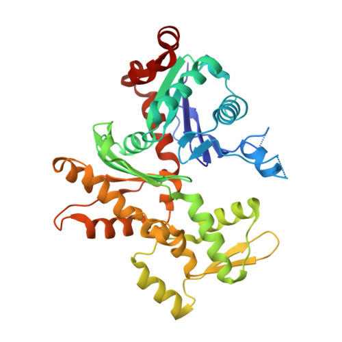
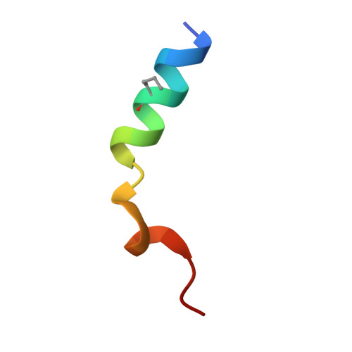
Structural evidence for the roles of divalent cations in actin polymerization and activation of ATP hydrolysis

Scipion, C.P.M.Ghoshdastider, U.Ferrer, F.J.Yuen, T.Y.Wongsantichon, J.Robinson, R.C.

(2018) Proc Natl Acad Sci U S A 115: 10345-10350

  • DOI: https://doi.org/10.1073/pnas.1806394115
  • Primary Citation of Related Structures:  
    5YPU

  • PubMed Abstract: 

    The structure of the actin filament is known at a resolution that has allowed the architecture of protein components to be unambiguously assigned. However, fully understanding the chemistry of the system requires higher resolution to identify the ions and water molecules involved in polymerization and ATP hydrolysis. Here, we find experimental evidence for the association of cations with the surfaces of G-actin in a 2.0-Å resolution X-ray structure of actin bound to a Cordon-Bleu WH2 motif and in previously determined high-resolution X-ray structures. Three of four reoccurring divalent cation sites were stable during molecular dynamics (MD) simulations of the filament, suggesting that these sites may play a functional role in stabilizing the filament. We modeled the water coordination at the ATP-bound Mg 2+ , which also proved to be stable during the MD simulations. Using this model of the filament with a hydrated ATP-bound Mg 2+ , we compared the cumulative probability of an activated hydrolytic water molecule approaching the γ-phosphorous of ATP, in comparison with G-actin, in the MD simulations. The cumulative probability increased in F-actin in line with the activation of actin's ATPase activity on polymerization. However, inclusion of the cations in the filament lowered cumulative probability, suggesting the rate of hydrolysis may be linked to filament flexibility. Together, these data extend the possible roles of Mg 2+ in polymerization and the mechanism of polymerization-induced activation of actin's ATPase activity.


  • Organizational Affiliation

    Institute of Molecular and Cell Biology, Agency for Science, Technology, and Research (A*STAR), Biopolis, 138673 Singapore.


Macromolecules
Find similar proteins by:  (by identity cutoff)  |  3D Structure
Entity ID: 1
MoleculeChains Sequence LengthOrganismDetailsImage
Actin, alpha skeletal muscle
A, C
368Oryctolagus cuniculusMutation(s): 0 
UniProt
Find proteins for P68135 (Oryctolagus cuniculus)
Explore P68135 
Go to UniProtKB:  P68135
Entity Groups  
Sequence Clusters30% Identity50% Identity70% Identity90% Identity95% Identity100% Identity
UniProt GroupP68135
Sequence Annotations
Expand
  • Reference Sequence

Find similar proteins by:  Sequence   |   3D Structure  

Entity ID: 2
MoleculeChains Sequence LengthOrganismDetailsImage
Cordon-Bleu WH2 motif
B, D
22Mus musculusMutation(s): 0 
UniProt
Find proteins for Q5NBX1 (Mus musculus)
Explore Q5NBX1 
Go to UniProtKB:  Q5NBX1
Entity Groups  
Sequence Clusters30% Identity50% Identity70% Identity90% Identity95% Identity100% Identity
UniProt GroupQ5NBX1
Sequence Annotations
Expand
  • Reference Sequence
Small Molecules
Modified Residues  1 Unique
IDChains TypeFormula2D DiagramParent
NLE
Query on NLE
B, D
L-PEPTIDE LINKINGC6 H13 N O2LEU
Experimental Data & Validation

Experimental Data

  • Method: X-RAY DIFFRACTION
  • Resolution: 2.00 Å
  • R-Value Free: 0.240 
  • R-Value Work: 0.193 
  • R-Value Observed: 0.195 
  • Space Group: C 1 2 1
Unit Cell:
Length ( Å )Angle ( ˚ )
a = 174.861α = 90
b = 40.832β = 101.76
c = 109γ = 90
Software Package:
Software NamePurpose
HKL-2000data collection
HKL-2000data scaling
REFMACrefinement
PDB_EXTRACTdata extraction
HKL-2000data reduction
MOLREPphasing

Structure Validation

View Full Validation Report


Ligand Structure Quality Assessment 


Entry History 

Deposition Data

Revision History  (Full details and data files)

  • Version 1.0: 2018-09-26
    Type: Initial release
  • Version 1.1: 2018-10-10
    Changes: Data collection, Database references, Structure summary
  • Version 1.2: 2018-10-24
    Changes: Data collection, Database references, Structure summary
  • Version 1.3: 2023-11-22
    Changes: Data collection, Database references, Derived calculations, Refinement description