5ZEO

X-ray structure of sperm whale V21C/V66C/F46S myoglobin mutant with an intramolecular disulfide bond


Experimental Data Snapshot

  • Method: X-RAY DIFFRACTION
  • Resolution: 1.77 Å
  • R-Value Free: 0.266 
  • R-Value Work: 0.223 

wwPDB Validation   3D Report Full Report


Ligand Structure Quality Assessment 


This is version 1.1 of the entry. See complete history


Literature

Regulation of both the structure and function by a de novo designed disulfide bond: a case study of heme proteins in myoglobin

Yin, L.L.Yuan, H.Du, K.J.He, B.Gao, S.Q.Wen, G.B.Tan, X.Lin, Y.W.

(2018) Chem Commun (Camb) 54: 4356-4359

  • DOI: https://doi.org/10.1039/c8cc01646a
  • Primary Citation of Related Structures:  
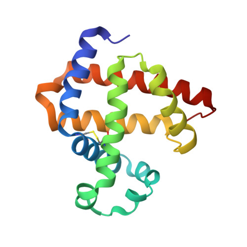
    5ZEO

  • PubMed Abstract: 

    A de novo designed intramolecular disulfide bond in myoglobin, resembling that in cytoglobin without structural evidence, was confirmed by an X-ray structure for the first time and was demonstrated to regulate both the structure and function of this protein, which fulfills the design of an artificial dehaloperoxidase, with an activity exceeding that of a native enzyme.


  • Organizational Affiliation

    School of Chemistry and Chemical Engineering, University of South China, Hengyang 421001, China. linlinying@hotmail.com ywlin@usc.edu.cn.


Macromolecules
Find similar proteins by:  (by identity cutoff)  |  3D Structure
Entity ID: 1
MoleculeChains Sequence LengthOrganismDetailsImage
Myoglobin153Physeter catodonMutation(s): 3 
Gene Names: MB
UniProt
Find proteins for P02185 (Physeter macrocephalus)
Explore P02185 
Go to UniProtKB:  P02185
Entity Groups  
Sequence Clusters30% Identity50% Identity70% Identity90% Identity95% Identity100% Identity
UniProt GroupP02185
Sequence Annotations
Expand
  • Reference Sequence
Small Molecules
Ligands 1 Unique
IDChains Name / Formula / InChI Key2D Diagram3D Interactions
HEM
Query on HEM

Download Ideal Coordinates CCD File 
B [auth A]PROTOPORPHYRIN IX CONTAINING FE
C34 H32 Fe N4 O4
KABFMIBPWCXCRK-RGGAHWMASA-L
Experimental Data & Validation

Experimental Data

  • Method: X-RAY DIFFRACTION
  • Resolution: 1.77 Å
  • R-Value Free: 0.266 
  • R-Value Work: 0.223 
  • Space Group: P 21 21 21
Unit Cell:
Length ( Å )Angle ( ˚ )
a = 39.566α = 90
b = 47.351β = 90
c = 79.111γ = 90
Software Package:
Software NamePurpose
REFMACrefinement
HKL-3000data reduction
HKL-3000data scaling
MOLREPphasing

Structure Validation

View Full Validation Report


Ligand Structure Quality Assessment 


Entry History 

Deposition Data

  • Released Date: 2018-05-09 
  • Deposition Author(s): Yuan, H.

Revision History  (Full details and data files)

  • Version 1.0: 2018-05-09
    Type: Initial release
  • Version 1.1: 2023-11-22
    Changes: Data collection, Database references, Refinement description