6BBN

Crystal structure of a curved tubulin complex induced by the kinesin-13 Kif2A


Experimental Data Snapshot

  • Method: X-RAY DIFFRACTION
  • Resolution: 3.51 Å
  • R-Value Free: 0.287 
  • R-Value Work: 0.236 
  • R-Value Observed: 0.239 

wwPDB Validation   3D Report Full Report


Ligand Structure Quality Assessment 


This is version 1.4 of the entry. See complete history


Literature

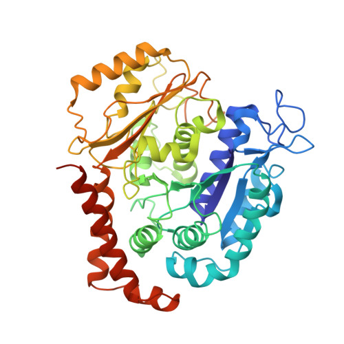
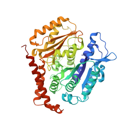
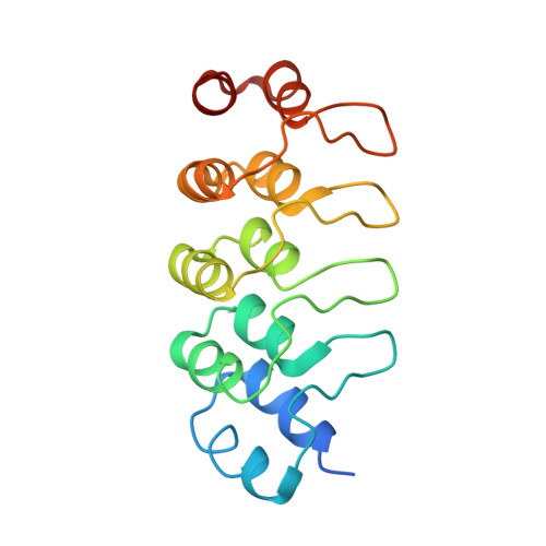
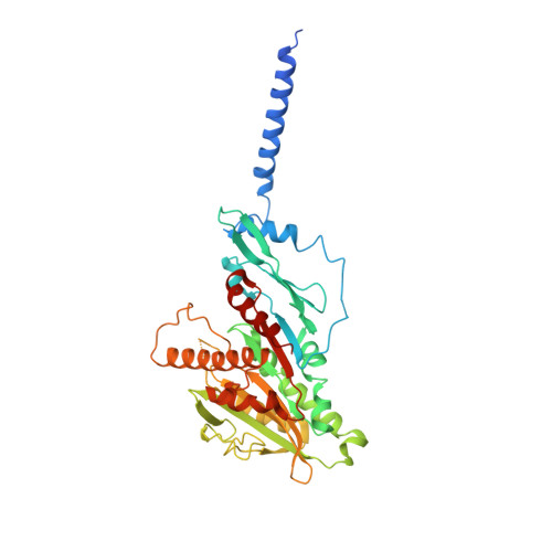
Ternary complex of Kif2A-bound tandem tubulin heterodimers represents a kinesin-13-mediated microtubule depolymerization reaction intermediate.

Trofimova, D.Paydar, M.Zara, A.Talje, L.Kwok, B.H.Allingham, J.S.

(2018) Nat Commun 9: 2628-2628

  • DOI: https://doi.org/10.1038/s41467-018-05025-7
  • Primary Citation of Related Structures:  
    6BBN

  • PubMed Abstract: 

    Kinesin-13 proteins are major microtubule (MT) regulatory factors that catalyze removal of tubulin subunits from MT ends. The class-specific "neck" and loop 2 regions of these motors are required for MT depolymerization, but their contributing roles are still unresolved because their interactions with MT ends have not been observed directly. Here we report the crystal structure of a catalytically active kinesin-13 monomer (Kif2A) in complex with two bent αβ-tubulin heterodimers in a head-to-tail array, providing a view of these interactions. The neck of Kif2A binds to one tubulin dimer and the motor core to the other, guiding insertion of the KVD motif of loop 2 in between them. AMPPNP-bound Kif2A can form stable complexes with tubulin in solution and trigger MT depolymerization. We also demonstrate the importance of the neck in modulating ATP turnover and catalytic depolymerization of MTs. These results provide mechanistic insights into the catalytic cycles of kinesin-13.


  • Organizational Affiliation

    Department of Biomedical and Molecular Sciences, Queen's University, Kingston, ON, K7L 3N6, Canada.


Macromolecules
Find similar proteins by:  (by identity cutoff)  |  3D Structure
Entity ID: 1
MoleculeChains Sequence LengthOrganismDetailsImage
Tubulin alpha-1B chain
A, C
451Bos taurusMutation(s): 0 
UniProt
Find proteins for P81947 (Bos taurus)
Explore P81947 
Go to UniProtKB:  P81947
Entity Groups  
Sequence Clusters30% Identity50% Identity70% Identity90% Identity95% Identity100% Identity
UniProt GroupP81947
Sequence Annotations
Expand
  • Reference Sequence
Find similar proteins by:  (by identity cutoff)  |  3D Structure
Entity ID: 2
MoleculeChains Sequence LengthOrganismDetailsImage
Tubulin beta-2B chain
B, D
445Bos taurusMutation(s): 0 
UniProt
Find proteins for Q6B856 (Bos taurus)
Explore Q6B856 
Go to UniProtKB:  Q6B856
Entity Groups  
Sequence Clusters30% Identity50% Identity70% Identity90% Identity95% Identity100% Identity
UniProt GroupQ6B856
Sequence Annotations
Expand
  • Reference Sequence
Find similar proteins by:  (by identity cutoff)  |  3D Structure
Entity ID: 3
MoleculeChains Sequence LengthOrganismDetailsImage
DARPinE [auth P]168Escherichia coliMutation(s): 0 
Entity Groups  
Sequence Clusters30% Identity50% Identity70% Identity90% Identity95% Identity100% Identity
Sequence Annotations
Expand
  • Reference Sequence
Find similar proteins by:  (by identity cutoff)  |  3D Structure
Entity ID: 4
MoleculeChains Sequence LengthOrganismDetailsImage
Kinesin-like protein KIF2AF [auth E]420Homo sapiensMutation(s): 0 
Gene Names: KIF2AKIF2KNS2
UniProt & NIH Common Fund Data Resources
Find proteins for O00139 (Homo sapiens)
Explore O00139 
Go to UniProtKB:  O00139
PHAROS:  O00139
GTEx:  ENSG00000068796 
Entity Groups  
Sequence Clusters30% Identity50% Identity70% Identity90% Identity95% Identity100% Identity
UniProt GroupO00139
Sequence Annotations
Expand
  • Reference Sequence
Small Molecules
Ligands 5 Unique
IDChains Name / Formula / InChI Key2D Diagram3D Interactions
GTP
Query on GTP

Download Ideal Coordinates CCD File 
G [auth A],
K [auth C]
GUANOSINE-5'-TRIPHOSPHATE
C10 H16 N5 O14 P3
XKMLYUALXHKNFT-UUOKFMHZSA-N
ANP
Query on ANP

Download Ideal Coordinates CCD File 
O [auth E]PHOSPHOAMINOPHOSPHONIC ACID-ADENYLATE ESTER
C10 H17 N6 O12 P3
PVKSNHVPLWYQGJ-KQYNXXCUSA-N
GDP
Query on GDP

Download Ideal Coordinates CCD File 
I [auth B],
M [auth D]
GUANOSINE-5'-DIPHOSPHATE
C10 H15 N5 O11 P2
QGWNDRXFNXRZMB-UUOKFMHZSA-N
PO4
Query on PO4

Download Ideal Coordinates CCD File 
N [auth D]PHOSPHATE ION
O4 P
NBIIXXVUZAFLBC-UHFFFAOYSA-K
MG
Query on MG

Download Ideal Coordinates CCD File 
H [auth A],
J [auth B],
L [auth C],
P [auth E]
MAGNESIUM ION
Mg
JLVVSXFLKOJNIY-UHFFFAOYSA-N
Experimental Data & Validation

Experimental Data

  • Method: X-RAY DIFFRACTION
  • Resolution: 3.51 Å
  • R-Value Free: 0.287 
  • R-Value Work: 0.236 
  • R-Value Observed: 0.239 
  • Space Group: P 1 21 1
Unit Cell:
Length ( Å )Angle ( ˚ )
a = 145.052α = 90
b = 82.188β = 109.33
c = 147.658γ = 90
Software Package:
Software NamePurpose
PHENIXrefinement
XSCALEdata scaling
PDB_EXTRACTdata extraction
XDSdata reduction
MOLREPphasing

Structure Validation

View Full Validation Report


Ligand Structure Quality Assessment 


Entry History & Funding Information

Deposition Data


Funding OrganizationLocationGrant Number
Canadian Institutes of Health Research (CIHR)Canada--
Natural Sciences and Engineering Research Council (NSERC, Canada)Canada--

Revision History  (Full details and data files)

  • Version 1.0: 2018-05-23
    Type: Initial release
  • Version 1.1: 2018-07-25
    Changes: Data collection, Database references
  • Version 1.2: 2019-02-20
    Changes: Author supporting evidence, Data collection
  • Version 1.3: 2020-01-08
    Changes: Author supporting evidence
  • Version 1.4: 2023-10-04
    Changes: Data collection, Database references, Derived calculations, Refinement description