6BBS

Joint X-ray/neutron structure of human carbonic anhydrase II in complex with brinzolamide


Experimental Data Snapshot

  • Method: X-RAY DIFFRACTION
  • Resolution: 2.00 Å
  • R-Value Free: 0.287 
  • R-Value Work: 0.276 

  • Method: NEUTRON DIFFRACTION

wwPDB Validation   3D Report Full Report


Ligand Structure Quality Assessment 


This is version 1.2 of the entry. See complete history


Literature

"To Be or Not to Be" Protonated: Atomic Details of Human Carbonic Anhydrase-Clinical Drug Complexes by Neutron Crystallography and Simulation.

Kovalevsky, A.Aggarwal, M.Velazquez, H.Cuneo, M.J.Blakeley, M.P.Weiss, K.L.Smith, J.C.Fisher, S.Z.McKenna, R.

(2018) Structure 26: 383-390.e3

  • DOI: https://doi.org/10.1016/j.str.2018.01.006
  • Primary Citation of Related Structures:  
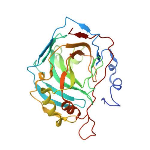
    6BBS, 6BC9, 6BCC

  • PubMed Abstract: 

    Human carbonic anhydrases (hCAs) play various roles in cells, and have been drug targets for decades. Sequence similarities of hCA isoforms necessitate designing specific inhibitors, which requires detailed structural information for hCA-inhibitor complexes. We present room temperature neutron structures of hCA II in complex with three clinical drugs that provide in-depth analysis of drug binding, including protonation states of the inhibitors, hydration water structure, and direct visualization of hydrogen-bonding networks in the enzyme's active site. All sulfonamide inhibitors studied bind to the Zn metal center in the deprotonated, anionic, form. Other chemical groups of the drugs can remain neutral or be protonated when bound to hCA II. MD simulations have shown that flexible functional groups of the inhibitors may alter their conformations at room temperature and occupy different sub-sites. This study offers insights into the design of specific drugs to target cancer-related hCA isoform IX.


  • Organizational Affiliation

    Biology and Soft Matter Division, Oak Ridge National Laboratory, Oak Ridge, TN 37831, USA. Electronic address: kovalevskyay@ornl.gov.


Macromolecules
Find similar proteins by:  (by identity cutoff)  |  3D Structure
Entity ID: 1
MoleculeChains Sequence LengthOrganismDetailsImage
Carbonic anhydrase 2260Homo sapiensMutation(s): 0 
Gene Names: CA2
EC: 4.2.1.1
UniProt & NIH Common Fund Data Resources
Find proteins for P00918 (Homo sapiens)
Explore P00918 
Go to UniProtKB:  P00918
PHAROS:  P00918
GTEx:  ENSG00000104267 
Entity Groups  
Sequence Clusters30% Identity50% Identity70% Identity90% Identity95% Identity100% Identity
UniProt GroupP00918
Sequence Annotations
Expand
  • Reference Sequence
Small Molecules
Ligands 2 Unique
IDChains Name / Formula / InChI Key2D Diagram3D Interactions
BZ1
Query on BZ1

Download Ideal Coordinates CCD File 
C [auth A](+)-4-ETHYLAMINO-3,4-DIHYDRO-2-(METHOXY)PROPYL-2H-THIENO[3,2-E]-1,2-THIAZINE-6-SULFONAMIDE-1,1-DIOXIDE
C12 H21 N3 O5 S3
HCRKCZRJWPKOAR-JTQLQIEISA-N
ZN
Query on ZN

Download Ideal Coordinates CCD File 
B [auth A]ZINC ION
Zn
PTFCDOFLOPIGGS-UHFFFAOYSA-N
Binding Affinity Annotations 
IDSourceBinding Affinity
BZ1 BindingDB:  6BBS Ki: min: 0.13, max: 45 (nM) from 6 assay(s)
Kd: min: 0.09, max: 0.13 (nM) from 3 assay(s)
IC50: min: 3.2, max: 45 (nM) from 2 assay(s)
Experimental Data & Validation

Experimental Data

  • Method: X-RAY DIFFRACTION
  • Resolution: 2.00 Å
  • R-Value Free: 0.287 
  • R-Value Work: 0.276 
  • Space Group: P 1 21 1
Unit Cell:
Length ( Å )Angle ( ˚ )
a = 44.755α = 90
b = 44.011β = 104.18
c = 75.957γ = 90
Software Package:
Software NamePurpose
nCNSrefinement

Structure Validation

View Full Validation Report


Ligand Structure Quality Assessment 


Entry History 

Deposition Data

Revision History  (Full details and data files)

  • Version 1.0: 2018-02-28
    Type: Initial release
  • Version 1.1: 2018-03-21
    Changes: Database references
  • Version 1.2: 2024-03-13
    Changes: Data collection, Database references