6BNV

CryoEM structure of MyosinVI-actin complex in the rigor (nucleotide-free) state, backbone-averaged with side chains truncated to alanine


Experimental Data Snapshot

  • Method: ELECTRON MICROSCOPY
  • Resolution: 4.60 Å
  • Aggregation State: FILAMENT 
  • Reconstruction Method: HELICAL 

wwPDB Validation   3D Report Full Report


This is version 1.2 of the entry. See complete history


Literature

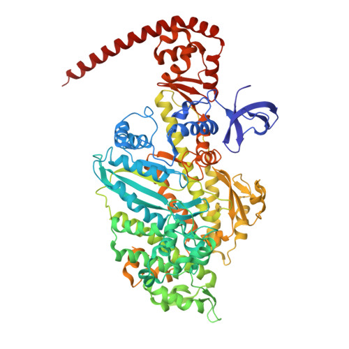
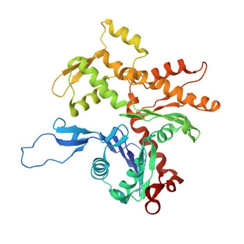
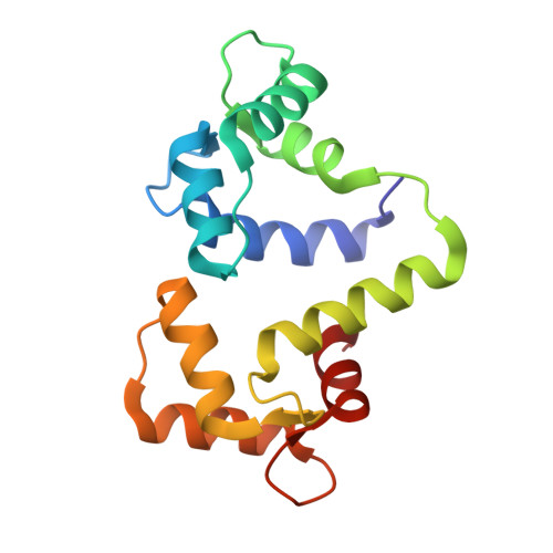
Cryo-EM structures reveal specialization at the myosin VI-actin interface and a mechanism of force sensitivity.

Gurel, P.S.Kim, L.Y.Ruijgrok, P.V.Omabegho, T.Bryant, Z.Alushin, G.M.

(2017) Elife 6

  • DOI: https://doi.org/10.7554/eLife.31125
  • Primary Citation of Related Structures:  
    6BNO, 6BNP, 6BNQ, 6BNU, 6BNV, 6BNW

  • PubMed Abstract: 

    Despite extensive scrutiny of the myosin superfamily, the lack of high-resolution structures of actin-bound states has prevented a complete description of its mechanochemical cycle and limited insight into how sequence and structural diversification of the motor domain gives rise to specialized functional properties. Here we present cryo-EM structures of the unique minus-end directed myosin VI motor domain in rigor (4.6 Å) and Mg-ADP (5.5 Å) states bound to F-actin. Comparison to the myosin IIC-F-actin rigor complex reveals an almost complete lack of conservation of residues at the actin-myosin interface despite preservation of the primary sequence regions composing it, suggesting an evolutionary path for motor specialization. Additionally, analysis of the transition from ADP to rigor provides a structural rationale for force sensitivity in this step of the mechanochemical cycle. Finally, we observe reciprocal rearrangements in actin and myosin accompanying the transition between these states, supporting a role for actin structural plasticity during force generation by myosin VI.


  • Organizational Affiliation

    Laboratory of Structural Biophysics and Mechanobiology, The Rockefeller University, New York, United States.


Macromolecules
Find similar proteins by:  (by identity cutoff)  |  3D Structure
Entity ID: 1
MoleculeChains Sequence LengthOrganismDetailsImage
Unconventional myosin-VI816Sus scrofaMutation(s): 0 
Gene Names: MYO6
UniProt
Find proteins for Q29122 (Sus scrofa)
Explore Q29122 
Go to UniProtKB:  Q29122
Entity Groups  
Sequence Clusters30% Identity50% Identity70% Identity90% Identity95% Identity100% Identity
UniProt GroupQ29122
Sequence Annotations
Expand
  • Reference Sequence
Find similar proteins by:  (by identity cutoff)  |  3D Structure
Entity ID: 2
MoleculeChains Sequence LengthOrganismDetailsImage
Actin, alpha skeletal muscle373Oryctolagus cuniculusMutation(s): 0 
UniProt
Find proteins for P68135 (Oryctolagus cuniculus)
Explore P68135 
Go to UniProtKB:  P68135
Entity Groups  
Sequence Clusters30% Identity50% Identity70% Identity90% Identity95% Identity100% Identity
UniProt GroupP68135
Sequence Annotations
Expand
  • Reference Sequence
Find similar proteins by:  (by identity cutoff)  |  3D Structure
Entity ID: 3
MoleculeChains Sequence LengthOrganismDetailsImage
Calmodulin
O, P, Q, R, S
O, P, Q, R, S, T
145Gallus gallusMutation(s): 0 
Gene Names: CALMCAMRCJMB04_24e7
UniProt
Find proteins for P62149 (Gallus gallus)
Explore P62149 
Go to UniProtKB:  P62149
Entity Groups  
Sequence Clusters30% Identity50% Identity70% Identity90% Identity95% Identity100% Identity
UniProt GroupP62149
Sequence Annotations
Expand
  • Reference Sequence
Experimental Data & Validation

Experimental Data

  • Method: ELECTRON MICROSCOPY
  • Resolution: 4.60 Å
  • Aggregation State: FILAMENT 
  • Reconstruction Method: HELICAL 
EM Software:
TaskSoftware PackageVersion
RECONSTRUCTIONFREALIGN9.11
MODEL REFINEMENTMDFF
MODEL REFINEMENTPHENIX

Structure Validation

View Full Validation Report


Entry History & Funding Information

Deposition Data


Funding OrganizationLocationGrant Number
National Institutes of Health/Office of the DirectorUnited States5DP5OD017885

Revision History  (Full details and data files)

  • Version 1.0: 2018-01-10
    Type: Initial release
  • Version 1.1: 2018-01-17
    Changes: Author supporting evidence
  • Version 1.2: 2024-03-13
    Changes: Data collection, Database references