6BQX

Crystal structure of Escherichia coli DsbA in complex with N-methyl-1-(4-phenoxyphenyl)methanamine


Experimental Data Snapshot

  • Method: X-RAY DIFFRACTION
  • Resolution: 1.99 Å
  • R-Value Free: 0.244 
  • R-Value Work: 0.176 
  • R-Value Observed: 0.183 

wwPDB Validation   3D Report Full Report


Ligand Structure Quality Assessment 


This is version 1.3 of the entry. See complete history

Re-refinement Note

This entry reflects an alternative modeling of the original data in: 4wf5


Literature

Inhibition of Diverse DsbA Enzymes in Multi-DsbA Encoding Pathogens.

Totsika, M.Vagenas, D.Paxman, J.J.Wang, G.Dhouib, R.Sharma, P.Martin, J.L.Scanlon, M.J.Heras, B.

(2018) Antioxid Redox Signal 29: 653-666

  • DOI: https://doi.org/10.1089/ars.2017.7104
  • Primary Citation of Related Structures:  
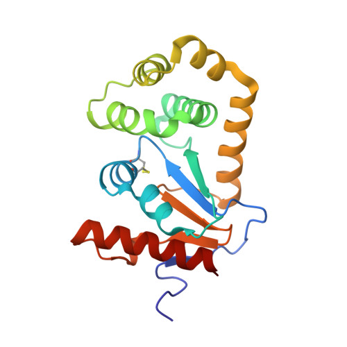
    6BQX, 6BR4

  • PubMed Abstract: 

    DsbA catalyzes disulfide bond formation in secreted and outer membrane proteins in bacteria. In pathogens, DsbA is a major facilitator of virulence constituting a target for antivirulence antimicrobial development. However, many pathogens encode multiple and diverse DsbA enzymes for virulence factor folding during infection. The aim of this study was to determine whether our recently identified inhibitors of Escherichia coli K-12 DsbA can inhibit the diverse DsbA enzymes found in two important human pathogens and attenuate their virulence.


  • Organizational Affiliation

    1 Institute of Health and Biomedical Innovation, School of Biomedical Sciences, Queensland University of Technology , Queensland, Australia .


Macromolecules
Find similar proteins by:  (by identity cutoff)  |  3D Structure
Entity ID: 1
MoleculeChains Sequence LengthOrganismDetailsImage
Thiol:disulfide interchange protein DsbA
A, B
189Escherichia coli K-12Mutation(s): 0 
Gene Names: dsbAdsfppfAb3860JW3832
UniProt
Find proteins for P0AEG4 (Escherichia coli (strain K12))
Explore P0AEG4 
Go to UniProtKB:  P0AEG4
Entity Groups  
Sequence Clusters30% Identity50% Identity70% Identity90% Identity95% Identity100% Identity
UniProt GroupP0AEG4
Sequence Annotations
Expand
  • Reference Sequence
Small Molecules
Ligands 1 Unique
IDChains Name / Formula / InChI Key2D Diagram3D Interactions
9AG
Query on 9AG

Download Ideal Coordinates CCD File 
C [auth A]N-methyl-1-(4-phenoxyphenyl)methanamine
C14 H15 N O
YWBKPTGYRUSBFV-UHFFFAOYSA-N
Experimental Data & Validation

Experimental Data

  • Method: X-RAY DIFFRACTION
  • Resolution: 1.99 Å
  • R-Value Free: 0.244 
  • R-Value Work: 0.176 
  • R-Value Observed: 0.183 
  • Space Group: C 1 2 1
Unit Cell:
Length ( Å )Angle ( ˚ )
a = 117.15α = 90
b = 64.901β = 126.53
c = 75.416γ = 90
Software Package:
Software NamePurpose
HKL-2000data reduction
HKL-2000data scaling
PHASERphasing
PHENIXrefinement
PDB_EXTRACTdata extraction

Structure Validation

View Full Validation Report


Ligand Structure Quality Assessment 


Entry History & Funding Information

Deposition Data


Funding OrganizationLocationGrant Number
Australian Research Council (ARC)AustraliaDP150102287
Australian Research Council (ARC)AustraliaFT130100580
Australian Synchrotron Research Program FellowshipAustralia--

Revision History  (Full details and data files)

  • Version 1.0: 2017-12-27
    Type: Initial release
  • Version 1.1: 2018-02-14
    Changes: Database references
  • Version 1.2: 2018-08-08
    Changes: Data collection, Database references
  • Version 1.3: 2020-01-01
    Changes: Author supporting evidence