6BYU

X-ray crystal structure of Escherichia coli RNA polymerase (RpoB-H526Y) and ppApp complex


Experimental Data Snapshot

  • Method: X-RAY DIFFRACTION
  • Resolution: 3.60 Å
  • R-Value Free: 0.311 
  • R-Value Work: 0.244 
  • R-Value Observed: 0.245 

wwPDB Validation   3D Report Full Report


Ligand Structure Quality Assessment 


This is version 2.2 of the entry. See complete history


Literature

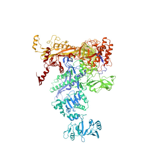
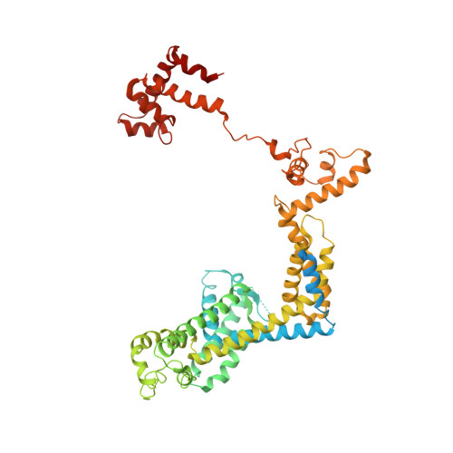
Structure-function comparisons of (p)ppApp vs (p)ppGpp for Escherichia coli RNA polymerase binding sites and for rrnB P1 promoter regulatory responses in vitro.

Bruhn-Olszewska, B.Molodtsov, V.Sobala, M.Dylewski, M.Murakami, K.S.Cashel, M.Potrykus, K.

(2018) Biochim Biophys Acta 1861: 731-742

  • DOI: https://doi.org/10.1016/j.bbagrm.2018.07.005
  • Primary Citation of Related Structures:  
    6BYU

  • PubMed Abstract: 

    Precise regulation of gene expression is crucial for bacteria to respond to changing environmental conditions. In addition to protein factors affecting RNA polymerase (RNAP) activity, second messengers play an important role in transcription regulation, such as well-known effectors of the stringent response: guanosine 5'triphosphate-3'diphosphate and guanosine 3', 5'-bis(diphosphate) [(p)ppGpp]. Although much is known about importance of the 5' and 3' moieties of (p)ppGpp, the role of the guanine base remains somewhat cryptic. Here, we use (p)ppGpp's adenine analogs [(p)ppApp] to investigate how the nucleobase contributes to determine its binding site and transcriptional regulation. We determined X-ray crystal structure of Escherichia coli RNAP-(p)ppApp complex, which shows the analogs bind near the active site and switch regions of RNAP. We have also explored the regulatory effects of (p)ppApp on transcription initiating from the well-studied E. coli rrnB P1 promoter to assess and compare properties of (p)ppApp with (p)ppGpp. We demonstrate that contrary to (p)ppGpp, (p)ppApp activates transcription at this promoter and DksA hinders this effect. Moreover, pppApp exerts a stronger effect than ppApp. We also show that when ppGpp and pppApp are present together, the outcome depends on which one of them was pre-incubated with RNAP first. This behavior suggests a surprising Yin-Yang like reciprocal plasticity of RNAP responses at a single promoter, occasioned simply by pre-exposure to one or the other nucleotide. Our observations underscore the importance of the (p)ppNpp's purine nucleobase for interactions with RNAP, which may lead to a better fundamental understanding of (p)ppGpp regulation of RNAP activity.


  • Organizational Affiliation

    Department of Bacterial Molecular Genetics, Faculty of Biology, University of Gdansk, Wita Stwosza 59, 80-308 Gdansk, Poland. Electronic address: b.bruhn.olszewska@ug.edu.pl.


Macromolecules
Find similar proteins by:  (by identity cutoff)  |  3D Structure
Entity ID: 1
MoleculeChains Sequence LengthOrganismDetailsImage
DNA-directed RNA polymerase subunit alpha
A, B, G, H
329Escherichia coli K-12Mutation(s): 0 
EC: 2.7.7.6
UniProt
Find proteins for P0A7Z4 (Escherichia coli (strain K12))
Explore P0A7Z4 
Go to UniProtKB:  P0A7Z4
Entity Groups  
Sequence Clusters30% Identity50% Identity70% Identity90% Identity95% Identity100% Identity
UniProt GroupP0A7Z4
Sequence Annotations
Expand
  • Reference Sequence
Find similar proteins by:  (by identity cutoff)  |  3D Structure
Entity ID: 2
MoleculeChains Sequence LengthOrganismDetailsImage
DNA-directed RNA polymerase subunit beta
C, I
1,342Escherichia coli K-12Mutation(s): 1 
EC: 2.7.7.6
UniProt
Find proteins for P0A8V2 (Escherichia coli (strain K12))
Explore P0A8V2 
Go to UniProtKB:  P0A8V2
Entity Groups  
Sequence Clusters30% Identity50% Identity70% Identity90% Identity95% Identity100% Identity
UniProt GroupP0A8V2
Sequence Annotations
Expand
  • Reference Sequence
Find similar proteins by:  (by identity cutoff)  |  3D Structure
Entity ID: 3
MoleculeChains Sequence LengthOrganismDetailsImage
DNA-directed RNA polymerase subunit beta'
D, J
1,407Escherichia coli K-12Mutation(s): 0 
EC: 2.7.7.6
UniProt
Find proteins for P0A8T7 (Escherichia coli (strain K12))
Explore P0A8T7 
Go to UniProtKB:  P0A8T7
Entity Groups  
Sequence Clusters30% Identity50% Identity70% Identity90% Identity95% Identity100% Identity
UniProt GroupP0A8T7
Sequence Annotations
Expand
  • Reference Sequence
Find similar proteins by:  (by identity cutoff)  |  3D Structure
Entity ID: 4
MoleculeChains Sequence LengthOrganismDetailsImage
DNA-directed RNA polymerase subunit omega
E, K
91Escherichia coli K-12Mutation(s): 0 
EC: 2.7.7.6
UniProt
Find proteins for P0A800 (Escherichia coli (strain K12))
Explore P0A800 
Go to UniProtKB:  P0A800
Entity Groups  
Sequence Clusters30% Identity50% Identity70% Identity90% Identity95% Identity100% Identity
UniProt GroupP0A800
Sequence Annotations
Expand
  • Reference Sequence
Find similar proteins by:  (by identity cutoff)  |  3D Structure
Entity ID: 5
MoleculeChains Sequence LengthOrganismDetailsImage
RNA polymerase sigma factor RpoD
F, L
613Escherichia coli K-12Mutation(s): 0 
UniProt
Find proteins for P00579 (Escherichia coli (strain K12))
Explore P00579 
Go to UniProtKB:  P00579
Entity Groups  
Sequence Clusters30% Identity50% Identity70% Identity90% Identity95% Identity100% Identity
UniProt GroupP00579
Sequence Annotations
Expand
  • Reference Sequence
Small Molecules
Ligands 3 Unique
IDChains Name / Formula / InChI Key2D Diagram3D Interactions
ECJ
Query on ECJ

Download Ideal Coordinates CCD File 
M [auth C],
Q [auth I]
(5R)-5-(6-amino-9H-purin-9-yl)-2-({[(S)-hydroxy(phosphonooxy)phosphoryl]oxy}methyl)-4-oxo-4,5-dihydrofuran-3-yl trihydrogen diphosphate
C10 H13 N5 O16 P4
CDOGMROMDFBAJR-SNVBAGLBSA-N
ZN
Query on ZN

Download Ideal Coordinates CCD File 
O [auth D],
P [auth D],
S [auth J],
T [auth J]
ZINC ION
Zn
PTFCDOFLOPIGGS-UHFFFAOYSA-N
MG
Query on MG

Download Ideal Coordinates CCD File 
N [auth D],
R [auth J]
MAGNESIUM ION
Mg
JLVVSXFLKOJNIY-UHFFFAOYSA-N
Experimental Data & Validation

Experimental Data

  • Method: X-RAY DIFFRACTION
  • Resolution: 3.60 Å
  • R-Value Free: 0.311 
  • R-Value Work: 0.244 
  • R-Value Observed: 0.245 
  • Space Group: P 21 21 21
Unit Cell:
Length ( Å )Angle ( ˚ )
a = 186.254α = 90
b = 203.656β = 90
c = 308.322γ = 90
Software Package:
Software NamePurpose
PHENIXrefinement
HKL-2000data reduction
HKL-2000data scaling
PHENIXphasing

Structure Validation

View Full Validation Report


Ligand Structure Quality Assessment 


Entry History & Funding Information

Deposition Data


Funding OrganizationLocationGrant Number
National Institutes of Health/National Institute of General Medical Sciences (NIH/NIGMS)United StatesGM087350

Revision History  (Full details and data files)

  • Version 1.0: 2018-01-17
    Type: Initial release
  • Version 1.1: 2018-02-14
    Changes: Author supporting evidence
  • Version 1.2: 2018-08-01
    Changes: Data collection, Database references
  • Version 1.3: 2018-08-15
    Changes: Data collection, Source and taxonomy, Structure summary
  • Version 2.0: 2018-08-29
    Changes: Atomic model, Data collection, Derived calculations, Source and taxonomy, Structure summary
  • Version 2.1: 2020-01-01
    Changes: Author supporting evidence
  • Version 2.2: 2023-10-04
    Changes: Data collection, Database references, Derived calculations, Refinement description