6BZ4

Human IgG1 lacking complement-dependent cytotoxicity: hu3S193 Fc mutant K322A


Experimental Data Snapshot

  • Method: X-RAY DIFFRACTION
  • Resolution: 2.40 Å
  • R-Value Free: 0.249 
  • R-Value Work: 0.212 
  • R-Value Observed: 0.216 

wwPDB Validation   3D Report Full Report


This is version 2.1 of the entry. See complete history


Literature

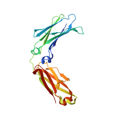
Global conformational changes in IgG-Fc upon mutation of the FcRn-binding site are not associated with altered antibody-dependent effector functions.

Burvenich, I.J.G.Farrugia, W.Liu, Z.Makris, D.King, D.Gloria, B.Perani, A.Allan, L.C.Scott, A.M.Ramsland, P.A.

(2018) Biochem J 475: 2179-2190

  • DOI: https://doi.org/10.1042/BCJ20180139
  • Primary Citation of Related Structures:  
    6BZ4

  • PubMed Abstract: 

    Antibody engineering is important for many diagnostic and clinical applications of monoclonal antibodies. We recently reported a series of fragment crystallizable (Fc) mutations targeting the neonatal Fc receptor (FcRn) site on a Lewis Y (Le y ) binding IgG1, hu3S193. The hu3S193 variants displayed shortened in vivo half-lives and may have potential for radioimaging or radiotherapy of Le y -positive tumors. Here, we report Fc crystal structures of wild-type hu3S193, seven FcRn-binding site variants, and a variant lacking C1q binding or complement-dependent cytotoxicity (CDC) activity. The Fc conformation of the FcRn-binding sites was similar for wild-type and all mutants of hu3S193 Fc, which suggests that FcRn interactions were directly affected by the amino acid substitutions. The C1q-binding site mutant Fc was nearly identical with the wild-type Fc. Surprisingly, several hu3S193 Fc variants showed large changes in global structure compared with wild-type Fc. All hu3S193 Fc mutants had similar antibody-dependent cellular cytotoxicity, despite some with conformations expected to diminish Fc gamma receptor binding. Several hu3S193 variants displayed altered CDC, but there was no correlation with the different Fc conformations. All versions of hu3S193, except the C1q-binding site mutant, bound C1q, suggesting that the altered CDC of some variants could result from different propensities to form IgG hexamers after engaging Le y on target cells. Overall, our findings support the concept that the antibody Fc is both flexible and mobile in solution. Structure-based design approaches should take into account the conformational plasticity of the Fc when engineering antibodies with optimal effector properties.


  • Organizational Affiliation

    Tumour Targeting Laboratory, Olivia Newton-John Cancer Research Institute, Melbourne, Australia.


Macromolecules
Find similar proteins by:  (by identity cutoff)  |  3D Structure
Entity ID: 1
MoleculeChains Sequence LengthOrganismDetailsImage
Immunoglobulin gamma-1 heavy chain
A, B
208Homo sapiensMutation(s): 1 
UniProt
Find proteins for P0DOX5 (Homo sapiens)
Explore P0DOX5 
Go to UniProtKB:  P0DOX5
Entity Groups  
Sequence Clusters30% Identity50% Identity70% Identity90% Identity95% Identity100% Identity
UniProt GroupP0DOX5
Sequence Annotations
Expand
  • Reference Sequence
Oligosaccharides

Help

Entity ID: 2
MoleculeChains Length2D Diagram Glycosylation3D Interactions
2-acetamido-2-deoxy-beta-D-glucopyranose-(1-2)-alpha-D-mannopyranose-(1-6)-[alpha-D-mannopyranose-(1-3)]beta-D-mannopyranose-(1-4)-2-acetamido-2-deoxy-beta-D-glucopyranose-(1-4)-[alpha-L-fucopyranose-(1-6)]2-acetamido-2-deoxy-beta-D-glucopyranose
C
7N-Glycosylation
Glycosylation Resources
GlyTouCan:  G45889JQ
GlyCosmos:  G45889JQ
GlyGen:  G45889JQ
Entity ID: 3
MoleculeChains Length2D Diagram Glycosylation3D Interactions
2-acetamido-2-deoxy-beta-D-glucopyranose-(1-2)-alpha-D-mannopyranose-(1-6)-[alpha-D-mannopyranose-(1-3)]beta-D-mannopyranose-(1-4)-2-acetamido-2-deoxy-beta-D-glucopyranose-(1-4)-2-acetamido-2-deoxy-beta-D-glucopyranose
D
6N-Glycosylation
Glycosylation Resources
GlyTouCan:  G64481DJ
GlyCosmos:  G64481DJ
GlyGen:  G64481DJ
Small Molecules
Ligands 1 Unique
IDChains Name / Formula / InChI Key2D Diagram3D Interactions
EDO
Query on EDO

Download Ideal Coordinates CCD File 
E [auth B]1,2-ETHANEDIOL
C2 H6 O2
LYCAIKOWRPUZTN-UHFFFAOYSA-N
Experimental Data & Validation

Experimental Data

  • Method: X-RAY DIFFRACTION
  • Resolution: 2.40 Å
  • R-Value Free: 0.249 
  • R-Value Work: 0.212 
  • R-Value Observed: 0.216 
  • Space Group: P 21 21 21
Unit Cell:
Length ( Å )Angle ( ˚ )
a = 49.324α = 90
b = 79.502β = 90
c = 138.221γ = 90
Software Package:
Software NamePurpose
PHENIXrefinement
XDSdata reduction
XDSdata scaling
PHENIXphasing

Structure Validation

View Full Validation Report


Entry History & Funding Information

Deposition Data


Funding OrganizationLocationGrant Number
National Health and Medical Research Council (NHMRC, Australia)Australia542512
National Health and Medical Research Council (NHMRC, Australia)Australia1030469

Revision History  (Full details and data files)

  • Version 1.0: 2018-06-13
    Type: Initial release
  • Version 1.1: 2018-07-18
    Changes: Data collection, Database references
  • Version 1.2: 2020-01-08
    Changes: Author supporting evidence, Data collection
  • Version 2.0: 2020-07-29
    Type: Remediation
    Reason: Carbohydrate remediation
    Changes: Atomic model, Data collection, Derived calculations, Structure summary
  • Version 2.1: 2023-10-04
    Changes: Data collection, Database references, Derived calculations, Refinement description, Structure summary