6CM3

BG505 SOSIP in complex with sCD4, 17b, 8ANC195


Experimental Data Snapshot

  • Method: ELECTRON MICROSCOPY
  • Resolution: 3.54 Å
  • Aggregation State: PARTICLE 
  • Reconstruction Method: SINGLE PARTICLE 

wwPDB Validation   3D Report Full Report


This is version 2.0 of the entry. See complete history


Literature

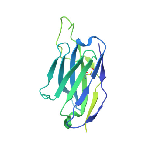
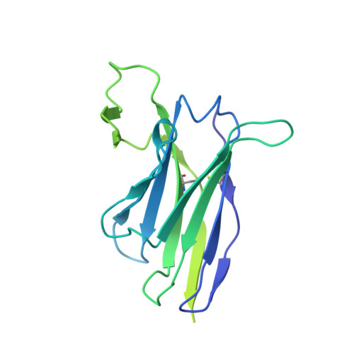
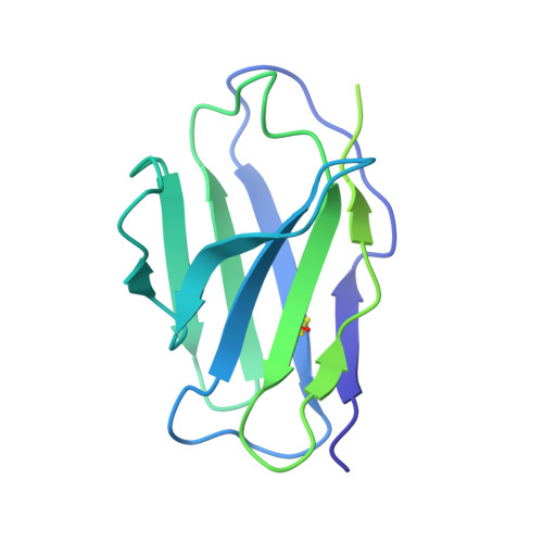
Partially Open HIV-1 Envelope Structures Exhibit Conformational Changes Relevant for Coreceptor Binding and Fusion.

Wang, H.Barnes, C.O.Yang, Z.Nussenzweig, M.C.Bjorkman, P.J.

(2018) Cell Host Microbe 24: 579-592.e4

  • DOI: https://doi.org/10.1016/j.chom.2018.09.003
  • Primary Citation of Related Structures:  
    6CM3, 6EDU

  • PubMed Abstract: 

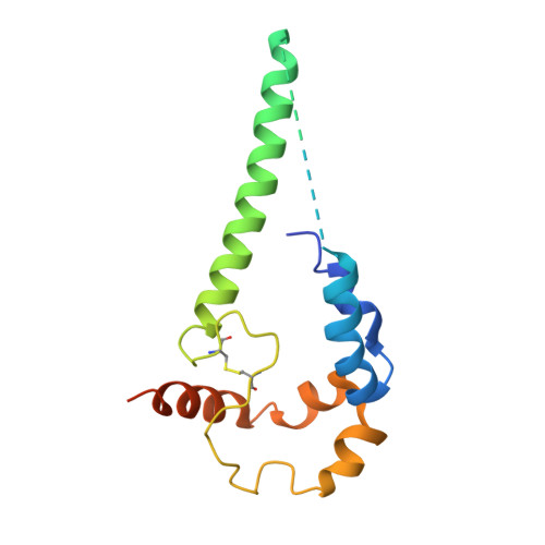
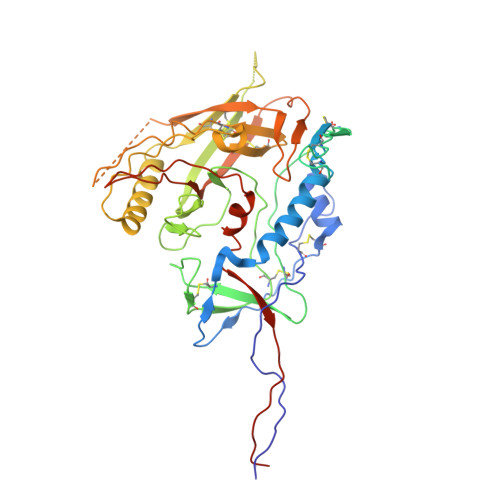
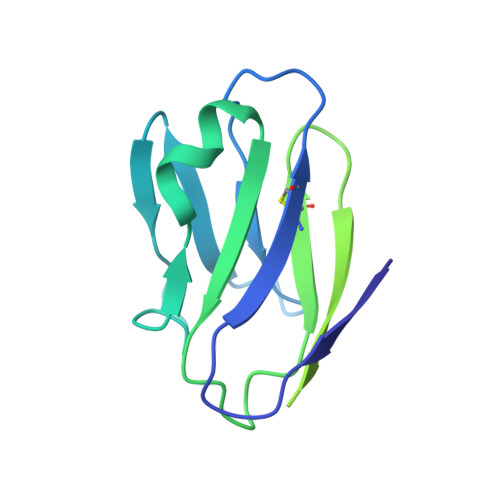
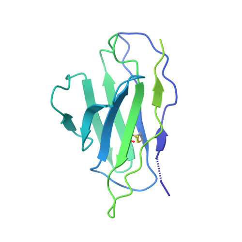
    HIV-1 Env, a trimer of gp120-gp41 heterodimers, mediates membrane fusion after binding host receptor CD4. Receptor binding displaces V1V2 loops from Env's apex, allowing coreceptor binding and opening Env to enable gp41-mediated fusion. We present 3.54 Å and 4.06 Å cryoelectron microscopy structures of partially open soluble native-like Env trimers (SOSIPs) bound to CD4. One structure, a complex with a coreceptor-mimicking antibody that binds both CD4 and gp120, stabilizes the displaced V1V2 and reveals its structure. Comparing partially and fully open Envs with closed Envs shows that gp41 rearrangements are independent of the CD4-induced rearrangements that result in V1V2 displacement and formation of a 4-stranded bridging sheet. These findings suggest ordered conformational changes before coreceptor binding: (1) gp120 opening inducing side-chain rearrangements and a compact gp41 central helix conformation, and (2) 4-stranded bridging-sheet formation and V1V2 displacement. These analyses illuminate potential receptor-induced Env changes and inform design of therapeutics disrupting viral entry.


  • Organizational Affiliation

    Division of Biology and Biological Engineering, California Institute of Technology, 1200 E. California Boulevard, Pasadena, CA 91125, USA.


Macromolecules
Find similar proteins by:  (by identity cutoff)  |  3D Structure
Entity ID: 1
MoleculeChains Sequence LengthOrganismDetailsImage
Envelope glycoprotein gp160
A, B, C
153Human immunodeficiency virus 1Mutation(s): 0 
Gene Names: env
UniProt
Find proteins for Q2N0S7 (Human immunodeficiency virus 1)
Explore Q2N0S7 
Go to UniProtKB:  Q2N0S7
Entity Groups  
Sequence Clusters30% Identity50% Identity70% Identity90% Identity95% Identity100% Identity
UniProt GroupQ2N0S7
Sequence Annotations
Expand
  • Reference Sequence
Find similar proteins by:  (by identity cutoff)  |  3D Structure
Entity ID: 2
MoleculeChains Sequence LengthOrganismDetailsImage
Envelope glycoprotein gp160
D, E, F
481Human immunodeficiency virus 1Mutation(s): 0 
Gene Names: env
UniProt
Find proteins for Q2N0S5 (Human immunodeficiency virus 1)
Explore Q2N0S5 
Go to UniProtKB:  Q2N0S5
Entity Groups  
Sequence Clusters30% Identity50% Identity70% Identity90% Identity95% Identity100% Identity
UniProt GroupQ2N0S5
Sequence Annotations
Expand
  • Reference Sequence
Find similar proteins by:  (by identity cutoff)  |  3D Structure
Entity ID: 3
MoleculeChains Sequence LengthOrganismDetailsImage
T-cell surface glycoprotein CD4
G, H, I
192Homo sapiensMutation(s): 0 
Gene Names: CD4
UniProt & NIH Common Fund Data Resources
Find proteins for P01730 (Homo sapiens)
Explore P01730 
Go to UniProtKB:  P01730
PHAROS:  P01730
GTEx:  ENSG00000010610 
Entity Groups  
Sequence Clusters30% Identity50% Identity70% Identity90% Identity95% Identity100% Identity
UniProt GroupP01730
Sequence Annotations
Expand
  • Reference Sequence
Find similar proteins by:  (by identity cutoff)  |  3D Structure
Entity ID: 4
MoleculeChains Sequence LengthOrganismDetailsImage
17b Fab light chain
J, L, N
214Homo sapiensMutation(s): 0 
Entity Groups  
Sequence Clusters30% Identity50% Identity70% Identity90% Identity95% Identity100% Identity
Sequence Annotations
Expand
  • Reference Sequence
Find similar proteins by:  (by identity cutoff)  |  3D Structure
Entity ID: 5
MoleculeChains Sequence LengthOrganismDetailsImage
17b Fab heavy chain
K, M, O
229Homo sapiensMutation(s): 0 
Entity Groups  
Sequence Clusters30% Identity50% Identity70% Identity90% Identity95% Identity100% Identity
Sequence Annotations
Expand
  • Reference Sequence
Find similar proteins by:  (by identity cutoff)  |  3D Structure
Entity ID: 6
MoleculeChains Sequence LengthOrganismDetailsImage
8ANC195 Fab heavy chain
P, R, T
244Homo sapiensMutation(s): 0 
Entity Groups  
Sequence Clusters30% Identity50% Identity70% Identity90% Identity95% Identity100% Identity
Sequence Annotations
Expand
  • Reference Sequence
Find similar proteins by:  (by identity cutoff)  |  3D Structure
Entity ID: 7
MoleculeChains Sequence LengthOrganismDetailsImage
8ANC195 Fab light chain
Q, S, U
215Homo sapiensMutation(s): 0 
Entity Groups  
Sequence Clusters30% Identity50% Identity70% Identity90% Identity95% Identity100% Identity
Sequence Annotations
Expand
  • Reference Sequence
Oligosaccharides

Help

Entity ID: 8
MoleculeChains Length2D Diagram Glycosylation3D Interactions
2-acetamido-2-deoxy-beta-D-glucopyranose-(1-4)-2-acetamido-2-deoxy-beta-D-glucopyranose
BA [auth b],
FA [auth f],
JA [auth j],
V,
W,
BA [auth b],
FA [auth f],
JA [auth j],
V,
W,
X
2N-Glycosylation
Glycosylation Resources
GlyTouCan:  G42666HT
GlyCosmos:  G42666HT
GlyGen:  G42666HT
Entity ID: 9
MoleculeChains Length2D Diagram Glycosylation3D Interactions
alpha-D-mannopyranose-(1-2)-alpha-D-mannopyranose-(1-2)-alpha-D-mannopyranose-(1-3)-[alpha-D-mannopyranose-(1-2)-alpha-D-mannopyranose-(1-6)-[alpha-D-mannopyranose-(1-3)]alpha-D-mannopyranose-(1-6)]beta-D-mannopyranose-(1-4)-2-acetamido-2-deoxy-beta-D-glucopyranose-(1-4)-2-acetamido-2-deoxy-beta-D-glucopyranoseCA [auth c],
GA [auth g],
Y
10N-Glycosylation
Glycosylation Resources
GlyTouCan:  G40702WU
GlyCosmos:  G40702WU
GlyGen:  G40702WU
Entity ID: 10
MoleculeChains Length2D Diagram Glycosylation3D Interactions
alpha-D-mannopyranose-(1-2)-alpha-D-mannopyranose-(1-3)-beta-D-mannopyranose-(1-4)-2-acetamido-2-deoxy-beta-D-glucopyranose-(1-4)-2-acetamido-2-deoxy-beta-D-glucopyranoseDA [auth d],
HA [auth h],
Z
5N-Glycosylation
Glycosylation Resources
GlyTouCan:  G42227JK
GlyCosmos:  G42227JK
GlyGen:  G42227JK
Entity ID: 11
MoleculeChains Length2D Diagram Glycosylation3D Interactions
alpha-D-mannopyranose-(1-3)-[alpha-D-mannopyranose-(1-6)]beta-D-mannopyranose-(1-4)-2-acetamido-2-deoxy-beta-D-glucopyranose-(1-4)-2-acetamido-2-deoxy-beta-D-glucopyranoseAA [auth a],
EA [auth e],
IA [auth i]
5N-Glycosylation
Glycosylation Resources
GlyTouCan:  G22768VO
GlyCosmos:  G22768VO
GlyGen:  G22768VO
Small Molecules
Ligands 1 Unique
IDChains Name / Formula / InChI Key2D Diagram3D Interactions
NAG
Query on NAG

Download Ideal Coordinates CCD File 
AB [auth F]
BB [auth F]
KA [auth D]
LA [auth D]
MA [auth D]
AB [auth F],
BB [auth F],
KA [auth D],
LA [auth D],
MA [auth D],
NA [auth D],
OA [auth D],
PA [auth D],
QA [auth E],
RA [auth E],
SA [auth E],
TA [auth E],
UA [auth E],
VA [auth E],
WA [auth F],
XA [auth F],
YA [auth F],
ZA [auth F]
2-acetamido-2-deoxy-beta-D-glucopyranose
C8 H15 N O6
OVRNDRQMDRJTHS-FMDGEEDCSA-N
Experimental Data & Validation

Experimental Data

  • Method: ELECTRON MICROSCOPY
  • Resolution: 3.54 Å
  • Aggregation State: PARTICLE 
  • Reconstruction Method: SINGLE PARTICLE 

Structure Validation

View Full Validation Report


Entry History & Funding Information

Deposition Data


Funding OrganizationLocationGrant Number
National Institutes of Health/National Institute Of Allergy and Infectious Diseases (NIH/NIAID)United StatesHIVRAD P01 AI100148

Revision History  (Full details and data files)

  • Version 1.0: 2018-10-17
    Type: Initial release
  • Version 1.1: 2018-10-24
    Changes: Data collection, Database references
  • Version 1.2: 2019-12-18
    Changes: Author supporting evidence, Other
  • Version 2.0: 2020-07-29
    Type: Remediation
    Reason: Carbohydrate remediation
    Changes: Advisory, Atomic model, Data collection, Derived calculations, Structure summary