6CSX

Single particles Cryo-EM structure of AcrB D407A associated with lipid bilayer at 3.0 Angstrom


Experimental Data Snapshot

  • Method: ELECTRON MICROSCOPY
  • Resolution: 3.00 Å
  • Aggregation State: PARTICLE 
  • Reconstruction Method: SINGLE PARTICLE 

wwPDB Validation   3D Report Full Report


This is version 1.3 of the entry. See complete history


Literature

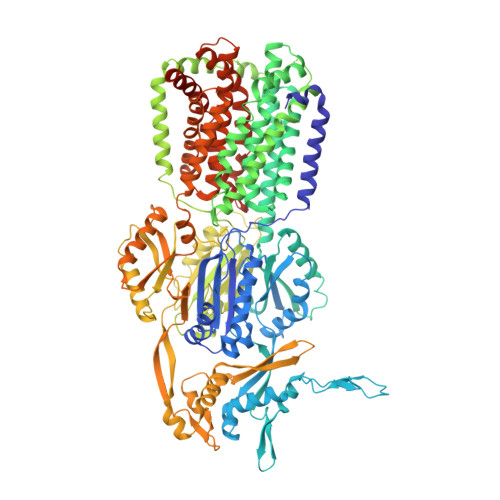
Structure and activity of lipid bilayer within a membrane-protein transporter.

Qiu, W.Fu, Z.Xu, G.G.Grassucci, R.A.Zhang, Y.Frank, J.Hendrickson, W.A.Guo, Y.

(2018) Proc Natl Acad Sci U S A 115: 12985-12990

  • DOI: https://doi.org/10.1073/pnas.1812526115
  • Primary Citation of Related Structures:  
    6BAJ, 6CSX

  • PubMed Abstract: 

    Membrane proteins function in native cell membranes, but extraction into isolated particles is needed for many biochemical and structural analyses. Commonly used detergent-extraction methods destroy naturally associated lipid bilayers. Here, we devised a detergent-free method for preparing cell-membrane nanoparticles to study the multidrug exporter AcrB, by cryo-EM at 3.2-Å resolution. We discovered a remarkably well-organized lipid-bilayer structure associated with transmembrane domains of the AcrB trimer. This bilayer patch comprises 24 lipid molecules; inner leaflet chains are packed in a hexagonal array, whereas the outer leaflet has highly irregular but ordered packing. Protein side chains interact with both leaflets and participate in the hexagonal pattern. We suggest that the lipid bilayer supports and harmonizes peristaltic motions through AcrB trimers. In AcrB D407A, a putative proton-relay mutant, lipid bilayer buttresses protein interactions lost in crystal structures after detergent-solubilization. Our detergent-free system preserves lipid-protein interactions for visualization and should be broadly applicable.


  • Organizational Affiliation

    Department of Medicinal Chemistry, Virginia Commonwealth University, Richmond, VA 23298.


Macromolecules
Find similar proteins by:  (by identity cutoff)  |  3D Structure
Entity ID: 1
MoleculeChains Sequence LengthOrganismDetailsImage
Multidrug efflux pump subunit AcrBA [auth B],
B [auth C],
C [auth A]
1,057Escherichia coli K-12Mutation(s): 1 
Gene Names: acrBacrEb0462JW0451
Membrane Entity: Yes 
UniProt
Find proteins for P31224 (Escherichia coli (strain K12))
Explore P31224 
Go to UniProtKB:  P31224
Entity Groups  
Sequence Clusters30% Identity50% Identity70% Identity90% Identity95% Identity100% Identity
UniProt GroupP31224
Sequence Annotations
Expand
  • Reference Sequence
Small Molecules
Ligands 2 Unique
IDChains Name / Formula / InChI Key2D Diagram3D Interactions
PTY
Query on PTY

Download Ideal Coordinates CCD File 
D [auth B]
E [auth B]
F [auth B]
G [auth B]
H [auth B]
D [auth B],
E [auth B],
F [auth B],
G [auth B],
H [auth B],
I [auth B],
J [auth B],
L [auth C],
M [auth C],
N [auth C],
O [auth C],
P [auth A],
Q [auth A],
R [auth A],
S [auth A],
T [auth A],
U [auth A],
V [auth A]
PHOSPHATIDYLETHANOLAMINE
C40 H80 N O8 P
NJGIRBISCGPRPF-KXQOOQHDSA-N
D12
Query on D12

Download Ideal Coordinates CCD File 
K [auth B]DODECANE
C12 H26
SNRUBQQJIBEYMU-UHFFFAOYSA-N
Experimental Data & Validation

Experimental Data

  • Method: ELECTRON MICROSCOPY
  • Resolution: 3.00 Å
  • Aggregation State: PARTICLE 
  • Reconstruction Method: SINGLE PARTICLE 

Structure Validation

View Full Validation Report


Entry History & Funding Information

Deposition Data


Funding OrganizationLocationGrant Number
Other governmentUnited StatesStartup

Revision History  (Full details and data files)

  • Version 1.0: 2018-12-05
    Type: Initial release
  • Version 1.1: 2018-12-19
    Changes: Data collection, Database references
  • Version 1.2: 2019-01-02
    Changes: Data collection, Database references
  • Version 1.3: 2024-03-13
    Changes: Data collection, Database references